Scientific Program
Keynote Session:
Title: One more year: Update on a longitudinal study of visual improvement and efficacy with transcorneal electrical stimulation retreatment in subjects with Retinitis Pigmentosa
Biography:
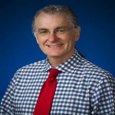
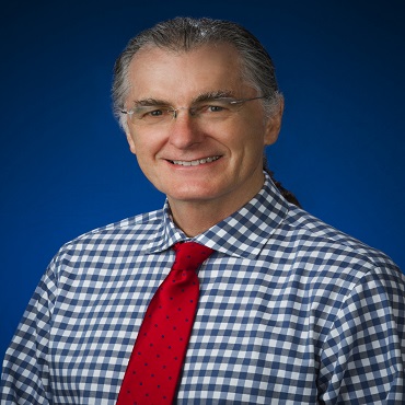
Kenneth R Seger received his doctorate of optometry from the University of California. Following a fellowship at AIIMS, he obtained an MSc from University of Manchester. He taught at the University of California, for 16 years. He has been at Nova Southeastern University for the last 20 years, teaching theoretical and practical classes as well as performing research. He has presented work in the USA, Europe and China. He is known for incorporating art, poetry and music into his courses. At NSU, he has been named professor of the year six times.
Abstract:
Some Retinitis Pigmentosa subjects show improved visual function following Transcorneal Electrical Stimulation (TES). The present study is a longitudinal report of the three subjects who were monitored greater than 41 months for declining visual function, which is expected in those with Retinitis Pigmentosa. The subjects received periodic retreatments of TES. Over a period of 41-43 months we monitored visual acuity, quick contrast sensitivity function and visual acuity. We found improvement in some of the functions for all three participants and did not see a drop below initial baseline in any of the subjects. Patient reports were consistent with the objective findings of enhanced visual performance. After encouraging visual improvements post TES that lasted several months, it appears restoration and/or prevention of slowly diminishing vision over time is possible with continued retreatments of TES. This requires large scale controlled study confirmation.
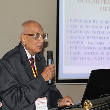
Title: A method of measuring eye injuries
Biography:
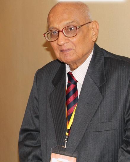
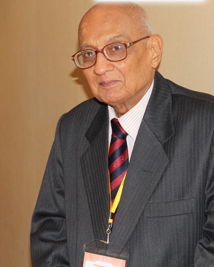
Bhartendu Shukla has teaching experience at medical college, gwalior for 25 years. Subsequently he was director, regional institute of ophthalmology, Bhopal for 9 years and now as director of research at R.J.N. for 9 years. He has been president at all India ophthalmological society & president, ocular trauma society of India. He received Dr. Siva Reddy International Award and important natioinal awards including Dr. Hari Mohan Award, Air Marshal Boparai Award, Community Ophthalmology Award, Dr. A.K.N. Sinha Award and Dr. Mohan Lal Award. He also received Life Time Achievements Award of All India Ophthalmological Society. He has over 50 publications in various national and international journals.
Abstract:
Measurement is the basis of scientific work and is done in a variety of ways. It is easy and accurate in physical sciences but not so in life sciences like medical science. A disease can be defined as a variable quantum of structural and or functional loss. Since the expression of these are very variable one can approximately convert the structural and functional loses in percentage and add them to get total loss. A method has been developed to do the same by making a traumagram on a graph paper. A horizontal straight line of 20 cm is drawn at the bottom of graph paper. From its centre a vertical 10 cm line is drawn which indicates the time period in weeks. Recording is started from the bottom line. On the left side structural loss is recorded and the functional loss on the right side by a point. This is recorded at weekly interval. Usually we a get a slanting line on either side. After 8-10 weeks the upper ends of both the lines are joined. This results in roughly a quadrangle figure which is termed traumagram. The mm squares enclosed in traumagram are counted and each is expressed as one trauma unit (1.T.U.). The total number of units indicate the magnitude of loss.
Title: Need of global parliament in teaching ophthalmology
Biography:
Katta S V has completed his MBBS, DO from S V University and fellow of retina foundation. He worked for 9 years under ministry of health, Algeria. He was senior resident in ophthalmology in Mallareddy medical college for women, India. He is maintaining retina eye clinic. He presented the grievance in general body AIOS 2015.
Abstract:
Article (226) high court & article (32) supreme court Indian penal code-applicable to all specialties. International court of justice only deals with land disputes. So, this presentation is needed to arrest further spread to younger generation.AIOS is second largest society in the world registered under society Act in the constitution of India. Are we justified in learning and also teaching knowingly non-precise incorrect medical terms to younger generation without even mentioning misnomers and in turn making them habituated? Under article 226 (high court) & 32 (Supreme Court) in the Constitution of India-Grievance. Our solution is to change or to teach misnomer along with the word. For the past 10 years, the same concept was being projected to the world as ‘Questionable medical terms in Ophthalmology’. As some of our AIOS governing council (2013-14) defended as lack of law and not authorized to opine on, retired judge was being consulted and the title is changed with law details as ‘Grievance in literature ophthalmology-our solutions’.Retrospective study of ophthalmic literature shows for the last more than hundred years, incorrect medical terms like ‘retinoscopy’, ‘syringing’, ‘retinal detachment’, ‘intracapsular cataract’ have been in existence. Phaco emulsification came approximately 28 years ago, 10 years ago small incision cataract surgery and 5 years ago computer vision syndrome. These less precise terms are being added to the medical literature during our own generation. No attempt has been made in the past even with appeal to restore precision and greater accuracy to the terminology in current use.
Title: Ocular injuries in children
Biography:
Bhartendu Shukla has teaching experience at medical college, gwalior for 25 years. Subsequently he was director, regional institute of ophthalmology, Bhopal for 9 years and now as director of research at R.J.N. for 9 years. He has been president at all India ophthalmological society & president, ocular trauma society of India. He received Dr. Siva Reddy International Award and important natioinal awards including Dr. Hari Mohan Award, Air Marshal Boparai Award, Community Ophthalmology Award, Dr. A.K.N. Sinha Award and Dr. Mohan Lal Award. He also received Life Time Achievements Award of All India Ophthalmological Society. He has over 50 publications in various national and international journals.
Abstract:
Ocular injuries are common and dangerous at all ages. However, they are more so in children for several reasons. In study of 1600 cases of ocular trauma 495 cases were in children(31%). In the sex ratio, male children were involved 3 times more. 60% were from the urban area and 40% from rural. In occupation 54% were students and 30% were nil as they were too young. The anterior segment was mainly involved. Maximum injuries occurred in the summer season(40%). Open globe injuries were slightly higher than closed globe injuries. Extraocular foreign bodies were double than the intraocular bodies. Earliest management is needed to prevent further damage.
Oral Session 1:
- Retina and Cornea Disorders | Cataract and Refractive surgery | Pediatric & Neuro Ophthalmology | Clinical and Surgical Ophthalmology | Nano Ophthalmology & Ophthalmic Drug Delivery

Chair
Kenneth R Seger
Nova Southeastern University, USA

Co-Chair
Bhartendu Shukla
R.J.N. Ophthalmic Institute, India
Title: Assessment of Intraocular pressure using Ocular Response Analyzer and Goldman Applanation Tonometry before and after Penetrating Keratoplasty (PKP)
Biography:
Wassef A, a young ophthalmologist, graduated medical school in 2014, completed Master degree in ophthalmology in May 2017 from Cairo University. working as an ophthalmology resident in Kasr Alainy medical school, Cairo university for three years and still. With special interest in cornea. Knowing that assessment of IOP post PKP poses a special challenge, motivated us to carry this research to test the different methods of assessment of IOP post PKP.
Abstract:
To compare the obtainability and values of IOP measurements by Goldmann Applanation Tonometry (GAT) and Ocular Response Analyzer (ORA) before and after penetrating keratoplasty (PKP) testing the degree of their agreement and the impact of corneal biomechanical factors on IOP measurement. Patients and Methods: The study is a comparative prospective study in which patients scheduled for penetrating keratoplasty (PKP) undergo intraocular pressure measurement (IOP) using the Ocular Response Analyzer (ORA) then the Goldman applanation tonometer (GAT) one day before surgery to be repeated one month after their surgery. Results: Forty patients undergoing PKP were enrolled in the study, 28 males (70%) and 12 females (30%). The mean age of patients involved is 42.8 ± 15.4, ranging between 9 and 75 years. Obtainability of ORA (92.5% of the patients) was significantly higher than GAT (60%) postoperatively (p<0.001), while no significant difference was elicited preoperatively. The mean cornea corrected IOP (IOPcc) was significantly higher than GAT and Goldmann related IOP (IOPg) both pre and postoperatively. In addition, both mean IOPcc and GAT postoperatively (19.2 ± 8.31, 15.65 ± 6.99 mmhg respectively) were significantly higher than their preoperative values (14.44 ± 7.03, 11.78 ± 4.55 mmhg respectively). Strong correlations existed between GAT and ORA measurements both pre and postoperatively. The level of agreement between GAT and IOPg was higher than IOPcc. Conclusion: ORA has proven to be superior to GAT in the ability to obtain reliable IOP measurements post PKP. IOPcc measurements also prooved to be relavant, independent on corneal biomechanical factors (CH and CRF) but judging the accuracy of its values needs further large scale studies.
Title: Rescuing retinal cells in vitro in age-related macular degeneration
Biography:
Sonali Nashine is an eye research scientist, her work is focused on retinal degeneration wherein the long-term goal is to identify therapeutic targets. During the course of research, she worked on models of retinal degenerative diseases, including retinitis pigmentosa, glaucoma, and age-related macular degeneration to identify candidate protective molecules. The ideal drugs will prolong the longevity of retinal cells, delay cell death, thereby saving vision.
Abstract:
Age-related macular degeneration is one of the primary causes of blindness in the United States as well as worldwide. The purpose of the present in vitro study was to test the hypothesis that natural compounds will rescue RPE cells from AMD mitochondria-induced damage, thereby rescuing AMD retinal cells. Our long-term goal is to develop a personalized treatment strategy for AMD patients. Herein, we treated AMD RPE transmitochondrial cells with naturally occurring antioxidants and neuroprotective compounds that are available as over-the-counter medications. AMD RPE transmitochondrial cells had identical nuclei but differed in mitochondrial DNA (mtDNA) content. Cell survival, free radical production, oxidative stress, and mitochondrial health was examined and compared between untreated AMD cells and the AMD cells treated with natural compounds. Treatment with natural compounds improved cell survival and mitochondrial health, reduced free radicals and oxidative stress, mitigated cell death, and increased live cell number in AMD cells compared to their untreated counterparts (p<0.05). In conclusion, the natural compounds used in this study might serve as effective, inexpensive, and non-invasive therapeutic options and might help in the development of a personalized treatment strategy for AMD patients.
Title: One More Year: Update on a longitudinal study of visual improvement and efficacy with transcorneal electrical stimulation retreatment in subjects with retinitis pigmentosa
Biography:
Kenneth R Seger received his doctorate of optometry from the University of California. Following a fellowship at All-India Institute of Medical Sciences, he obtained an MSc from University of Manchester. He taught at the University of California school of Optometry for 16 years. He has been at Nova Southeastern University for the last 20 years, teaching theoretical and practical classes as well as performing research.He has presented work in the USA, Europe and China.Dr. Seger is known for incorporating art, poetry and music into his courses. At NSU, he has been named Professor of the year six times.
Abstract:
Some Retinitis Pigmentosa (RP) subjects show improved visual function following Transcorneal Electrical Stimulation (TES). (1, 3) The present study is a longitudinal report of the three subjects who were monitored > 41 months for declining visual function, which is expected in those with RP. The subjects received periodic retreatments of TES. Over a period of 41-43 months we monitored visual acuity (VA), quick contrast sensitivity function (qCSF) and visual acuity (VA). We found improvement in some of the functions for all three participants and did not see a drop below initial baseline in any of the subjects. Patient reports were consistent with the objective findings of enhanced visual performance. After encouraging visual improvements post TES that lasted several months, it appears restoration and/or prevention of slowly diminishing vision (or, at least maintenance of baseline findings) over time is possible with continued retreatments of TES. This requires large scale controlled study confirmation.
Title: A Method of Measuring Eye Injuries
Biography:
Prof. B.Shukla has teaching experience at Medical Cololege,Gwalior for 25 years. Susequently he was Director, Regional Institute of Ophthalmology, Bhopoal for 9 years and now as Director of Research at R.J.N. for 9 years. He has been President All India OphthalmologicalSociety& President, Ocular Trauma Society of India. He received Dr. Siva Reddy International Award and important natioinal awards including Dr.Hari Mohan Award, Air Marshal Boparai Award, Community Ophthalmology Award, Dr.A.K.N. Sinha Award and Dr. Mohan Lal Award. He also receiuved Life Time Achievements Award of All India Ophthalmological Society. Dr.Shukla has over 50 publications in various national and international journals.
Abstract:
Measurement is the basis of scientific work and is done in a variety of ways. It is easy and accurate in physical sciences but not so in life sciences like medical science. A disease can be defined as a variable quantum of structural and or functional loss. Since the expression of these are very variable one can approximately convert the structural and functional loses in percentage and add them to get total loss. A method has been developed to do the same by making a traumagram on a graph paper. A horizontal straight line of 20 cm is drawn at the bottom of graph paper. From its centre a vertical 10 cm line is drawn which indicates the time period in weeks. Recording is started from the bottom line. On the left side structural loss is recorded and the functional loss on the right side by a point. This is recorded at weekly interval. Usually we a get a slanting line on either side. After 8-10 weeks the upper ends of both the lines are joined. This results in roughly a quadrangle figure which is termed traumagram. The mm squares enclosed in traumagram are counted and each is expressed as one trauma unit (1.T.U.). The total number of units indicate the magnitude of loss.
Title: Decoding and localising voluntary gaze abnormalities based upon clinical examination in Multiple Sclerosis
Biography:
Amar Kulkarni is a consultant ophthalmologist and phaco surgeon working in Doctor Eye Institute, India. He has completed his ophthalmology from Mysore Medical College and MM Joshi Eye Institute, India. He is an expert phaco surgeon who has performed more than 4000 cataract surgeries and handles all kinds of complicated cases. He is a very quick surgeon and expert in handling mass cases. His other areas of work include neuro-ophthalmology and medical retina. He is a fellow of the International Council of Ophthalmology. He has presented papers in state conferences and has published papers in international and national journals. He is very passionate about his work and likes to keep himself updated regularly.
Abstract:
Multiple Sclerosis (MS) is a demyelinating disease of the central nervous system leading to disability, especially in young patients. Acute or chronic lesions of MS within the brainstem and the cerebellum frequently results in ocular motor diseases. Eye movement disorders have the potential of being utilised as structural and functional biomarkers of early cognitive deficit and possibly help in assessing disease status and progression. Early detection of gaze palsies aids in diagnosis of various demyelinating conditions. This is also an important aspect of follow up examination as relapse, chronicity can be diagnosed with careful examination of gaze movements. The proper identification of voluntary gaze abnormality on clinical examination allows to map out site of demyelination and make neuroimaging precise and easier.
Title: Rescuing retinal cells in vitro in Age-Related Macular Degeneration(ARMD)
Biography:
Sonali Nashine is an eye research scientist, her work is focused on retinal degeneration wherein the long-term goal is to identify therapeutic targets. During the course of research, she worked on models of retinal degenerative diseases, including retinitis pigmentosa, glaucoma, and age-related macular degeneration to identify candidate protective molecules. The ideal drugs will prolong the longevity of retinal cells, delay cell death, thereby saving vision.
Abstract:
Age-related macular degeneration is one of the primary causes of blindness in the United States as well as worldwide. The purpose of the present in vitro study was to test the hypothesis that natural compounds will rescue RPE cells from AMD mitochondria-induced damage, thereby rescuing AMD retinal cells. Our long-term goal is to develop a personalized treatment strategy for AMD patients. Herein, we treated AMD RPE transmitochondrial cells with naturally occurring antioxidants and neuroprotective compounds that are available as over-the-counter medications. AMD RPE transmitochondrial cells had identical nuclei but differed in mitochondrial DNA (mtDNA) content. Cell survival, free radical production, oxidative stress, and mitochondrial health was examined and compared between untreated AMD cells and the AMD cells treated with natural compounds. Treatment with natural compounds improved cell survival and mitochondrial health, reduced free radicals and oxidative stress, mitigated cell death, and increased live cell number in AMD cells compared to their untreated counterparts (p<0.05). In conclusion, the natural compounds used in this study might serve as effective, inexpensive, and non-invasive therapeutic options and might help in the development of a personalized treatment strategy for AMD patients.
Title: Intravitreal anti-VEGF monotherapy in retinopathy of prematurity management, a case series of our first 10 eyes
Biography:
Rachid Chigara is ophthalmology trainee at Mustaphaan University hospital in Algiers. In his final year he has developed an interest in pediatric ophthalmology and currently works in the retinoblastoma unit at the same hospital, where conservative treatment of retinoblastoma is performed. After the Algerian society of ophthalmology’s 31st congress in 2016, whose main theme was ROP helped in retinopathy of prematurity screening and the treatment of certain cases using Anti-VEGF agents under the guidance of the head of the ophthalmology department.
Abstract:
The purpose is to study the structural outcome of intravitreal injections of Ranibizumab in zone 2 stage 3 ROP. Premature infants born in CHU-Mustapha maternity hospital, who met our main screening criteria, (Infants born before 30weeks gestational age and/or weighing less than 1500g at birth and/or required any sort of ventilation assistance and oxygen therapy) were automatically sent for screening following discharge. All screenings were carried out using a Retcam 3, after pupil dilation, using both tropicamide and phenylephrine, on awake infants. stage 3 zone 2 ROP were treated in the operating theatre under strict aseptic conditions with a single injection of 0.20mg Ranibizumab. Follow up exams were also carried out using a Retcam 3, after pupil dilation, on awake infants, 48 hours, days 7, 14, 21, and 28 as well as every second week for 3 months and every month for six months or until completion of retinal vascularisation.
Title: Assessment of intraocular pressure using ocular response Analyzer and goldman applanation tonometry before and after Penetrating Kerato Plasty (PKP)
Biography:
Wassef A, an young ophthalmologist, graduated medical school in 2014, completed Master degree (M.sc.) in ophthalmology in May 2017 from Cairo University. Working as an ophthalmology resident in Kasr Alainy medical school, Cairo University for three years and still with special interest in cornea knowing that assessment of IOP post PKP poses a special challenge, motivated all to carry this research to test the different methods of assessment of IOP post PKP.
Abstract:
To compare the obtainability and values of IOP measurements by Goldmann Applanation Tonometry (GAT) and Ocular Response Analyzer (ORA) before and after penetrating keratoplasty (PKP) testing the degree of their agreement and the impact of corneal biomechanical factors on IOP measurement. Patients and Methods: The study is a comparative prospective study in which patients scheduled for penetrating keratoplasty (PKP) undergo intraocular pressure measurement (IOP) using the Ocular Response Analyzer (ORA) then the Goldman applanation tonometer (GAT) one day before surgery to be repeated one month after their surgery. Results: Forty patients undergoing PKP were enrolled in the study, 28 males (70%) and 12 females (30%). The mean age of patients involved is 42.8 ± 15.4, ranging between 9 and 75 years. Obtainability of ORA (92.5% of the patients) was significantly higher than GAT (60%) postoperatively (p<0.001), while no significant difference was elicited preoperatively. The mean cornea corrected IOP (IOPcc) was significantly higher than GAT and Goldmann related IOP (IOPg) both pre and postoperatively. In addition, both mean IOPcc and GAT postoperatively (19.2 ± 8.31, 15.65 ± 6.99 mmhg respectively) were significantly higher than their preoperative values (14.44 ± 7.03, 11.78 ± 4.55 mmhg respectively). Strong correlations existed between GAT and ORA measurements both pre and postoperatively. The level of agreement between GAT and IOPg was higher than IOPcc. Conclusion: ORA has proven to be superior to GAT in the ability to obtain reliable IOP measurements post PKP. IOPcc measurements also prooved to be relavant, independent on corneal biomechanical factors (CH and CRF) but judging the accuracy of its values needs further large scale studies.
Title: Conformational and thermodynamic features of meibum in adolescents and adults with graft-versus-host disease
Biography:
Samiyyah Sledge received her master’s in Physiology and Biophysics from the University of Louisville School of Medicine just last year. Currently she is a doctoral candidate in the department of physiology and she researches in the department of ophthalmology and visual sciences under the guidance of Dr. Douglas Borchman. He has published at least five papers in several well regarded journals.
Abstract:
Graft-versus-host disease (GvHD) is a common complication of allogenic hematological stem cell transplantations that is often associated with dry eye disease (DED) with mixed Meibomian gland dysfunction and aqueous tear deficiency. The temperature-dependence of conformational changes in human meibum (hM) was measured in a comparative study to elucidate the potential impact of GvHD on hM composition, structure and function with DED. A total of 28 donors without DED and 15 donors with DED separated into adolescent and adult pools, were analyzed by Fourier Transform Infrared Spectroscopy. Both age and DED lead to conformational and thermodynamic differences in hM. Increases in lipid order and transition temperatures were observed in hM with GvHD. However, these relative increases were more pronounced in hM from adolescents than from adults. The decrease of change in enthalpy for adolescents was more pronounced compared with adults. The conformational and thermodynamic differences observed with age and, importantly, with DED/GvHD are indicative of compositional changes in hM that could impact tear film stability. We decided to test this theory. We hypothesize that the hM layer alone or hM layer interacting with phospholipids (PLs) in the aqueous portion of tears act as a barrier to tear evaporation; and that in patients with DED/GvHD, either altered composition of the hM and/or its interaction with PLs in tears, lead to a greater rate of evaporative fluid loss from tears. Our studies suggest that the hM alone does not retard the evaporation of water from a saline solution used as an aqueous phase.
Title: Innovative progressive lenses and study of limitations
Biography:
Katta S V has completed his MBBS, DO from S V University and fellow of retina foundation. He worked for 9 years under ministry of health, Algeria. He was senior resident in ophthalmology in Mallareddy medical college for women, India. He is maintaining retina eye clinic. He presented the grievance in general body AIOS 2015.
Abstract:
History of innovation, progressive of power causes fixed distance object visual strain because of inability to fix the visual gaze for longer periods. 10020 patients were studied in a period of 2 years and the results are all patients of fixed distant object viewers were unhappy with progressive lenses because of inability to fix visual gaze for longer periods. Conclusion is more than 1 dioptre addition advisable to take bifocals. Computer operators advisable to take near vision glasses with less of +0.25d.
Title: Need of Global parliament in teaching ophthalmology
Biography:
Katta S Venkateswar has completed his MBBS, DO from SV University and fellow of retina foundation. He worked for 9 years under ministry of health, Algeria. He was senior resident in ophthalmology in Mallareddy medical college for women, India. He is maintaining retina eye clinic. He presented the grievance in general body AIOS2015.
Abstract:
Article (226) high court & article (32) supreme court – Indian penal code-applicable to all specialties. International court of justice only deals with land disputes. So, this presentation is needed to arrest further spread to younger generation.AIOS is second largest society in the world registered under society Act in the constitution of India. Are we justified in learning and also teaching knowingly non-precise / incorrect medical terms to younger generation without even mentioning misnomers and in turn making them habituated? Under article 226 (high court) & 32 (Supreme Court) in the Constitution of India-Grievance. Our solution is to change or to teach misnomer along with the word. For the past 10 years, the same concept was being projected to the world as ‘Questionable medical terms in Ophthalmology’. As some of our AIOS governing council (2013-14) defended as lack of law and not authorized to opine on, retired judge was being consulted and the title is changed with law details as ‘Grievance in literature Ophthalmology-our solutions’.Retrospective study of ophthalmic literature shows for the last more than hundred years, incorrect medical terms like ‘retinoscopy’, ‘syringing’, ‘retinal detachment’, ‘intracapsular cataract’ have been in existence. Phaco emulsification came approximately 28 years ago, 10 years ago small incision cataract surgery and 5 years ago computer vision syndrome. These less precise terms are being added to the medical literature during our own generation. No attempt has been made in the past even with appeal to restore precision and greater accuracy to the terminology in current use. Perhaps none took cognizance of the problem or the need for such correction was not taken seriously. It is unethical that evidence based incorrect medical terms are being published in all scientific journals. Even with appeal, knowingly publishing incorrect medical terms in scientific journals and passing on to innocent younger generation is grievance. At present evidence based science is kept on the desk of chairman, ethics committee, international council of ophthalmology for consideration after relooking. Law is applied to all sciences. Ophthalmology is the starter of the procedure. Global parliament in Ophthalmology is needed in this concern. Search you tube: Ophthalmic visionary to all disciplines and Google search grievance in teaching ophthalmology.
Title: How to manage a case of hot orbit?
Biography:
Mohamed A Eldesouky has completed his PhD at the age of 36 years from Tanta University and Postdoctoral Studies from School of Medicine, University of Tennessee, Memphis. He is the Director of oculoplasty, and ocular pathology service in Tanta University Eye Hospital. He has published more than 25 papers in reputed journals.
Abstract:
Hot orbit is a term applied to a group of orbital disorders presented with the cardinal manifestations of inflammations as pain, redness, hotness and impaired ocular motility. This group of orbital disorders which varies from trivial as orbital hematoma to a very sever life and sometimes life threatening disorders as orbital metastasis and fungal orbital infections. In this study we describe our protocol in Tanta University Hospital Ophthalmology department, Egypt, for how to manage a case of hot orbit. In this study we present a tentative novel classification of hot orbit basically into infectious, non-infectious and mascaraed groups.

