

International Conference on
Medical Imaging and Clinical Research
Singapore City, Singapore February 25-26, 2019
Medical Imaging 2019
Theme: Medical Imaging for Improved Diagnosis and Treatment
With great pleasure we are inviting you to the upcoming Medical Imaging Conference 2019 that focus on the theme: "Medical Imaging for Improved Diagnosis and Treatment" scheduled in Singapore City, Singapore during February 25-26, 2019. Medical Imaging Congress gives a phase to globalize the investigation by presenting a talk among endeavors, educational affiliations and taking in return from research to industry. Medical Imaging and Clinical Research Conference focuses in communicating data and offers new considerations among the specialists, industrialists, and understudies from different domains of Medical Imaging to share their investigation experiences and appreciate savvy trades and novel sessions at the event. At Medical Imaging Meeting you can increase new information which will be outstandingly significant for development in the field and creating new plans and thoughts to improve yourself and your master calling. The field of Medical Imaging in diagnostics have not quite recently helped the headway in different fields in science and advancement yet, moreover, contributed towards the difference in the idea of human life in a manner of speaking. This has ended up being possible with the various disclosures and advancements advancing the change of various applications.
Medical Imaging Conference | Therapeutic Conference | Medical Imaging Meeting | Clinical Research Conference | Medical Imaging Events | Medical Imaging Events | Clinical Research Events | Medical Imaging Workshop | Clinical Research Workshop | Restorative Imaging Conference
Session-1: Medical Imaging
Medicinal Imaging is the strategy and procedure of making visual portrayals of the inside of a body for clinical investigation and therapeutic mediation and in addition visual portrayal of the capacity of a few organs or tissues. Medicinal imaging tries to uncover interior structures covered up by the skin and bones, and in addition to analyze and treat the ailment. Therapeutic imaging additionally builds up a database of typical life systems and physiology to make it conceivable to distinguish variations from the norm. In spite of the fact that imaging of expelled organs and tissues can be performed for therapeutic reasons, such methods are generally considered a piece of pathology rather than restorative imaging.
Medical Imaging Conference | Therapeutic Conference | Medical Imaging Meeting | Clinical Research Conference | Medical Imaging Events | Medical Imaging Events | Clinical Research Events | Medical Imaging Workshop | Clinical Research Workshop | Restorative Imaging Conference
Session-2: Therapeutic Imaging
Therapeutic imaging eludes to considerable measure diverse advancements that are utilized to see the human body so as to investigate, screen, or treat medical conditions. Medical imaging constitutes a database of normal anatomy and physiology to make it desirable to identify abnormalities. This includes the use of a variety of modalities, some of which may involve a risk of harmful ionizing emission. Due to the brisk advances in imaging technology, such as the introduction of multi-detector arrays and fast MRI protocols, both the number and variety of radiological applications are hysterically increasing. Each kind of innovation gives distinctive data about the territory of the body being considered or treated, identified with conceivable infection, damage, or the viability of therapeutic treatment.
Medical Imaging Conference | Therapeutic Conference | Medical Imaging Meeting | Clinical Research Conference | Medical Imaging Events | Medical Imaging Events | Clinical Research Events | Medical Imaging Workshop | Clinical Research Workshop | Restorative Imaging Conference
Session-3: Ultrasound Imaging
It uses high-frequency sound waves to view inside the body. Because ultrasound images are captured in real-time, they can also show the migration of the body's internal organs as well as blood flowing through the blood vessels. Unlike X-ray imaging, there is no ionizing radiation exposure associated with ultrasound imaging. This is commonly correlated with imaging the fetus in pregnant women. The ultrasound image is produced based on the reflection of the waves off of the body frame. The power of the sound signal and the time it takes for the wave to travel through the body provide the information necessary to produce an image.
Medical Imaging Conference | Therapeutic Conference | Medical Imaging Meeting | Clinical Research Conference | Medical Imaging Events | Medical Imaging Events | Clinical Research Events | Medical Imaging Workshop | Clinical Research Workshop | Restorative Imaging Conference
Session-4: Magnetic Resonance Imaging
MRI is a therapeutic imaging strategy for making images of the interior structures of the body. MRI scanners use strong magnetic fields and radio waves to make images. It uses powerful magnets to demarcate and excite hydrogen nuclei of water molecules in human tissue, producing an appreciable signal which is spatially encoded, resulting in images of the body. Like CT, MRI habitually creates a 2D image of a thin "slice" of the body and is therefore studied a tomographic imaging technique. Contemporary MRI instruments are capable of producing images in the form of 3D blocks, which may be considered a generalization of the single-slice, tomographic, concept. Amid an MRI exam, an electric current is gone through curled wires to make a brief attractive field in a patient's body. Radio waves are sent from and got by a transmitter/recipient in the machine, and these signs are utilized to make computerized pictures of the filtered zone of the body. MRI scans last from 20 - 90 minutes, depending on the part of the body being imaged.
Medical Imaging Conference | Therapeutic Conference | Medical Imaging Meeting | Clinical Research Conference | Medical Imaging Events | Medical Imaging Events | Clinical Research Events | Medical Imaging Workshop | Clinical Research Workshop | Restorative Imaging Conference
Session-5: Pediatric X-ray Imaging
Restorative x-beam imaging exams, which incorporate Computed tomography (CT), fluoroscopy, and traditional X-beams, utilize the most reduced radiation dosage obligatory, considering the size and age of the patient. It needs committed imaging conventions to procure the pictures, there is a requirement for acclimated anesthesia for profound techniques, for example, MRI, particular preparing is required for the medicinal services unit included, and exact information and skill ought to be connected for figuring the pictures. It requires consideration for radiation exposure if ionizing radiation is being used. One of the challenges for clinical care personnel is to gain the child's trust and co-operation before and throughout the duration of an examination, which can prove to be difficult in children who may be ill and have pain. This is important to acquire quality images and prevent repeat examinations.
Medical Imaging Conference | Therapeutic Conference | Medical Imaging Meeting | Clinical Research Conference | Medical Imaging Events | Medical Imaging Events | Clinical Research Events | Medical Imaging Workshop | Clinical Research Workshop | Restorative Imaging Conference
Session-6: Radiography
It is an imaging approach using X-rays to view the internal structure of an object. To construct the image, a beam of X-rays, a form of electromagnetic radiation, are composed by an X-ray alternator and are projected toward the object. A convinced amount of X-ray is absorbed by the object, defenseless on its density and composition.
The X-rays that pass through the object are conquering behind the object by a detector. Two forms of radiographic images are used Projection radiography and fluoroscopy. Fluoroscopy delivers ongoing pictures of inside structures of the body, however, draws in a consistent contribution of x-beams, at a lower measurements rate. Projection radiographs, regularly known as x-beams, are frequently used to control the sort and span of a break and in addition for identifying obsessive changes in the lungs.
Medical Imaging Conference | Therapeutic Conference | Medical Imaging Meeting | Clinical Research Conference | Medical Imaging Events | Medical Imaging Events | Clinical Research Events | Medical Imaging Workshop | Clinical Research Workshop | Restorative Imaging Conference
Session-7: Computed Tomography
It is also frequently referred to as a CAT scan, is a Medical Imaging process that combines multiple X-ray projections taken from distinct angles to produce detailed cross-sectional images of areas indoors the body. CT images allow doctors to get very decisive, 3-D views of convinced parts of the body, like as soft tissues, the pelvis, blood vessels, the lungs, the brain, the heart, abdomen, and bones. CT is also generally the preferred method of analyzing many cancers, such as liver, lung and pancreatic cancers. Digital geometry processing is used to farther provoke a three-dimensional volume of the inside of the object from an extensive sequence of two-dimensional radiographic images taken around a single axis of rotation. It has all the more as of late been utilized for a preventive solution or screening for an ailment, for instance, CT colonography for individuals with a high danger of colon tumor, or full-movement heart checks for individuals with a high danger of coronary illness.
Medical Imaging Conference | Therapeutic Conference | Medical Imaging Meeting | Clinical Research Conference | Medical Imaging Events | Medical Imaging Events | Clinical Research Events | Medical Imaging Workshop | Clinical Research Workshop | Restorative Imaging Conference
Session-8: Optical Imaging
Diffuse optical Imaging is a method of designing using near-infrared spectroscopy or fluorescence-established methods. When used to conceive 3D volumetric models of the imaged material DOI is referred to as diffuse optical tomography, though 2D imaging methods are confidential as diffuse optical topography. The disadvantage of optical imaging is the inadequacy of penetration depth, exclusively when working at visible wavelengths. The depth of penetration is linked to the absorption and scattering of light, which is primarily a function of the wavelength of the excitation source. Light is absorbed by endogenous chromophores found in living tissue. Examples include optical microscopy, spectroscopy, endoscopy, scanning laser ophthalmoscopy, and optical coherence tomography.
Medical Imaging Conference | Therapeutic Conference | Medical Imaging Meeting | Clinical Research Conference | Medical Imaging Events | Medical Imaging Events | Clinical Research Events | Medical Imaging Workshop | Clinical Research Workshop | Restorative Imaging Conference
Session-9: Near Infrared Imaging
Close Infrared Spectroscopy and Imaging utilizes nearby infrared light in the vicinity of 650 and 950 nm to non-obtrusively test the solidification and oxygenation of hemoglobin in the cerebrum, muscle alongside different tissues and is worn e.g. to identify changes persuaded by mind action, damage, or sickness. In brain analysis it complements functional magnetic resonance imaging (fMRI) by implementing measures of both oxygenated and deoxygenated haemoglobin concentrations and by permissive studies in populations of subjects with experimental paradigms that are not amenable to fMRI. Various close infrared (NIR) fluorophores have been utilized for in vivo imaging, including Kodak X-SIGHT Dyes and Conjugates, Pz 247, DyLight 750 and 800 Fluors, Although NIRS is commonly performed adopting instruments that emitted endless wave light and commonly measure the intensity of light inseminate through the tissue, it is also desirable to perform measurements where the source of light is intensity inflected (between 50 to 500 MHz) or oscillate (typically pulsed on for less than 100 ps) and the detector resolves correspondingly the phase or temporal delay of the light propagating over the tissue. These measurements are usually termed frequency domain or time domain measurements and because they provide direct measurements of photon propagation delay within the tissue as well as the intensity
Medical Imaging Conference | Therapeutic Conference | Medical Imaging Meeting | Clinical Research Conference | Medical Imaging Events | Medical Imaging Events | Clinical Research Events | Medical Imaging Workshop | Clinical Research Workshop | Restorative Imaging Conference
Session-10: Oncology Clinical Research
Medical Imaging and Clinical Research Conference aims at felicitating the meaningful discussion on modalities of improvement, contact sustenance,and care are used inside routine illness care to address and upgrade signs and individual fulfillment. Clinical Research Conference gathers professionals to discuss various ways challenges of paraneoplastic issue, or from treatment of harm that require provoke thought and reversal.
Medical Imaging Conference | Therapeutic Conference | Medical Imaging Meeting | Clinical Research Conference | Medical Imaging Events | Medical Imaging Events | Clinical Research Events | Medical Imaging Workshop | Clinical Research Workshop | Restorative Imaging Conference
The worldwide medical imaging market measure was esteemed USD 33.7 billion of every 2016 and is required to develop at a CAGR of 5.7% over the estimated time frame. Main considerations driving development of this industry is expanding interest for beginning period conclusion of ceaseless infection and rising maturing socioeconomics, which is required to support the request of symptomatic imaging over the globe.
The report considers the worldwide analytic imaging market over the estimated time of 2016 to 2021. The market is relied upon to achieve ~USD 36.43 Billion by 2021, at a CAGR of 6.6% from 2016 to 2021. Various factors, for example, expanding speculations, finances, and allows by government bodies for modernization of imaging offices; expanding ventures from open private associations; development in the quantity of indicative imaging focuses; rising pervasiveness of tumor; expanding geriatric populace and the resulting development in the rate of different infections; innovative headways in analytic imaging modalities; and expanding inclination for insignificantly intrusive medications drive the development of this market.
In any case, factors, for example, the high cost of demonstrative imaging frameworks, innovative confinements related with independent frameworks, negative human services changes in the U.S., and the deficiency of helium are relied upon to limit the development of this market to a specific degree.
Why Singapore?
Singapore’s healthcare system is the envy of the west, with the following factors being a key-factor for its efficient and affordable model: Public-Private Balance, Sustainable Financing, Strong Regulatory Governance. A system this good can serve as a benchmark for other countries, to improve successful implementation of policies and managing the economics as well.
In Singapore, more and more students are taking up clinical research as their career. The Singapore Ministry of Health states that the number of registrants for technicians courses are on the upraise. To encourage more people to take up clinical researchers, the government is also offering scholarships for students.
Currently, there are 29,894 Registered Medical Education Centers in Singapore, yet reports suggest that the country’s number of long-term care providers must be increased by 45% by 2020, to meet the requirements of the ageing population (The Lien Foundation, July 2018).
Industry Insights
Based on item, the market is fragmented into X-beam imaging frameworks, Computed tomography scanners, ultrasound imaging frameworks, Magnetic Resonance Imaging, and atomic imaging frameworks. Every methodology is additionally separated into sub fragments. The X-beam and ultrasound frameworks advertise is partitioned based on innovation and transportability; though, CT scanners are portioned by cut sort. X-ray frameworks are isolated based on engineering and field quality and the atomic imaging frameworks showcase is classified into SPECT and Hybrid PET frameworks. These frameworks are additionally partitioned into independent and half breed modalities.
In view of utilization, the market is fragmented into obstetrics/gynecology well being, orthopedics and musculoskeletal, neurology and spine, cardiovascular and thoracic, general imaging, bosom well being, and others. In view of end client, the market is fragmented into doctor's facilities, indicative imaging focuses, and opposite end clients (counting pharmaceutical and biotechnology organizations, scholastic and research focuses, sports foundations, and CROs).
U.S. medical imaging market, by product, 2014 - 2025 (USD Billion)
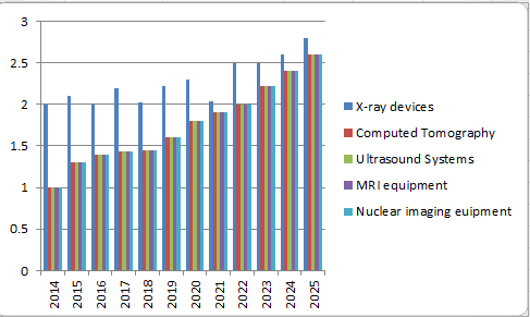
Product Insights
The medical imaging market is classified on the basis of product into X-ray, Computed Tomography, ultrasound system, MRI equipment, and nuclear imaging. The X-ray segment held the largest market share in 2016 and is expected to maintain its dominance over the forecast period.
This can be attributed to the increasing prevalence of orthopaedic injuries and accidents and the demand for point of care testing, which is facilitating the sale of portable devices. The X-ray devices are further segmented into stationary, and portable (handheld, and mobile).
The nuclear imaging segment is expected to be the fastest growing due to the development of new radiotracers, raising prevalence of cancer and cardiovascular diseases, and the introduction of new products through innovation and advancement. According to the American Cancer Society, Surveillance Research, there were about 1,665,540 cases of cancer in 2014, 1,685,210 cases of cancer in 2016.
Global medical imaging market, by application, 2016 (%)
Amid 2016, the general X-beam fragment was one of the quickest developing item portions in the worldwide therapeutic imaging market and will keep on dominating the market in the coming years. This fragment represents the most elevated income amid the estimate time frame as contrasted and different strategies, regardless of being the customary technique for therapeutic imaging. The progressions in innovation and expanded appropriation of compact X-beam machines will impel this present section's development prospects until the finish of 2021.
North America is the biggest market inferable from the appropriation of cutting edge framework and mindfulness about mechanical progressions. The Asia Pacific is anticipated to be the quickest developing business sector in light of the ascent in the geriatric populace, repayment approaches and tolerant mindfulness level in rising nations like India, China, Malaysia, and the Philippines.
A portion of the key players in worldwide Medical Imaging market are Mindray Medical International, Alpinion Medical Systems, BenQ Medical Technology, Boston Scientific, Carestream Health Inc, Esaote SpA, Fujifilm Holding, GE Healthcare, Hitachi Medical Corporation, Konica Minolta, Philips Healthcare, Shimadzu Corporation, Siemens Healthcare, Sonosite Inc and Toshiba Medical Systems.
Products Covered:
- Computed Tomography Scanners
- Portable/Mobile
- Stationary
- By Technology
- Low Slice Scanners
- Medium Slice Scanners
- High Slice Scanners
- Mammography Systems
- Analog Mammography Systems
- Digital Mammography Systems
- MRI Systems
- By Field Strength
- Low-to-Mid-Field MRI Systems
- High- & Very-High-Field MRI Systems
- Ultra-High-Field MRI Systems
- Closed MRI Systems
- Open MRI Systems
- Nuclear Imaging Systems/Radionuclide
- Single-Photon Emission Computed Tomography (SPECT) Scanners
- Position Emission Tomography (PET) Scanners
- Ultrasound Systems
- 2D
- 3D & 4D
- Doppler
- High Intensity Focused Ultrasound (HIFU)
- Extracorporeal Shockwave Lithotripsy (ESWL)
- By Portability
- Cart/Trolley Based
- Compact/Portable
- X-ray Imaging Systems
- Analog Imaging Systems
- Digital Imaging Systems
- By Portability
- Portable X-ray Imaging Systems
- Stationary X-ray Devices
- Tactile Imaging
- Thermography
- Elastography
Universities in Singapore:
Yong Loo Lin School of Medicine, Singapore
Academy of Medicine, Singapore
Duke-NUS Graduate School of Medicine, Singapore
John Hopkins International Medical centre, Singapore
Organization Associated in the USA:
American Association of Physicists in Medicine, USA
American Association for Women Radiologists, USA
American Board of Radiology, USA
American Brachytherapy Society, USA
American College of Medical Physics, USA
American College of Nuclear Medicine, USA
American College of Nuclear Physicians, USA
American College of Radiology, USA
Colombian Association of Radiology, USA
American College of Radiation Oncology, USA
Association of Educators in Imaging and Radiologic Sciences, USA
The Association for Medical Imaging Management, USA
Australian Institute of Radiography, USA
American Institute of Ultrasound in Medicine, USA
Mexico Association of Ultrasound in Medicine, USA
American Osteopathic College of Radiology, USA
American Physical Society, USA
Asia Pacific Society of Cardiovascular & Interventional Radiology, USA
American Registry of Diagnostic Medical Sonographers, USA
Association for Radiologic and Imaging, USA
American Roentgen Ray Society, USA
American Registry of Radiologic Technologists, USA
Institutes in the UK:
British Institute of Radiology, UK
British Nuclear Medicine Society, UK
British Society of Interventional Radiology, UK
British Society of Neuroradiologists, UK
Institutes in Canada:
Canadian Association of Medical Radiation Technologists, Canada
Canadian Association of Radiologists/L'Association canadienne des radiologists, Canada
Canadian Association of Radiation Oncologists/Association Canadienne des Radio-Oncologues, Canada
Canadian College of Physicists in Medicine, Canada
Computerized Medical Imaging Society, Canada
Institutes in Japan:
The University of Tokyo, Japan
Kyoto University, Japan
Osaka University, Japan
Tohoku University, Japan
Keio University, Japan
Nagoya University, Japan
Institutes in South Korea
Yonsei Universiy, South Korea
The University of Ulsan, South Korea
Sungkyun University, South Korea
Wonkwang University, South Korea
Soonchunhyang University, South Korea
- Medical Imaging and its Application
- Therapeutic Imaging
- Ultrasound Imaging
- Molecular Imaging, Functional imaging and Integrated Therapy
- Magnetic Resonance Imaging
- Radiography
- Computed Tomography
- Optical Imaging
- Oncology Clinical Research
- Journal of Clinical & Experimental Radiology
- Archives of Medical Biotechnology
- Journal of Clinical Images and Case Reports
8 Organizing Committee Members
Mohd Fahmi Mohd Yusof
Senior Lecturer
Universiti Sains Malaysia
Malaysia
Bhanu Prakash KN
Doctor
Singapore Bioimaging Consortium
Singapore
Alejandro De la Parra Solomon
CEO
Regener Age Clinic
Mexico
Joel I. Osorio
CEO
RegenerAge Clinic and RegenerAge Beauty
USA
Zang-Hee Cho
Professor
Seoul National University
Korea
Alireza Heidari
Prof
California South University (CSU), Irvine, California, USA
USA
Panayiotis Zavos
Professor
Andrology Institute of America
USA
Vikas Leelavati Balasaheb Jadhav
Consultant Radiologist
D.Y.Patil University
India
3 Renowned Speakers
Arathy John
Manipal Academy of Higher Education,
India
Keerthana Wilson
Assistant Professor and Echocardiographer
India
Kalaivaane Shanmugnathan
Mahasa University College
Malaysia
Media Partners & Collaborations
Media Partner

Media Partner

Media Partner

fgsg
tgh

Dear Eliza, Thank you for that wonderful opportunity given to share my experiences during the conference. I would like to place on record the smooth conduct of the conference. My appreciation goes a long way to Eliza for her timely communication and well advanced information disseminated processes. The presentation schedule and the time mentioned in the mail sent were also strictly followed. All the best. Regards Kalyani Kenneth Speaker | Psychiatry-2021
Kalyani Kenneth

Dear Laura, Thank you for giving me the opportunity to present my current work! I really enjoyed listening and connecting to a wider international scientist through this conference. And I am really looking forward for the physical conference next year! Best regards Sharmaine
Sharmaine Reintar

Many thanks Laura. It iş my plesure work with you
Pinar Kara

It was my pleasure to be a part of the conference. The talks were technically profound. Thank you once again.
Anupam Mukherjee

Thank you very much for the message. I am glad that the presented poster was appreciated. I'm also grateful for the certificate. Dispite my main work was the subject presented in the session, it would be a pleasure to participate in the webinars.
Ana Rita Domingues

Thank you for your appreciation. I love the support received from you and your admirable patience from invitation till the end of the conference. Thank you very much. I hope to participate in future conferences.
Munezza Khan

Am really grateful for you inviting me to this great event... Am thrilled to be part of this great team, I feel honored 😀😃😄🤝🏻
Aysha Haruna

The conference was arranged nicely through a Webex. I found the sessions interactive and informative. I am looking forward to join the next annual meeting of plant genomics in osaka, japan in april 2022.
Ruchika

Highly appreciate your quick response and I am grateful for all your help.
Dr. P T Sunderam

Informative and Innovative sessions
Michael Lacroix

Delightful event with a wonderful sessions
Mohamed Ebraheem Elmesserey

Very much grateful for this opportunity to share the innovative ideas
Mahmoud Metwaly Taha

It was really informative and excellent platform to share the research ideas
Raktima Chakrabarti

Wonderful experience
KHALED NASR ELDIN REYAD

Thank you very much for organizing this wonderful conference
JIMMY KAYASTHA

it was impressive and left me a very good memory.
VAISHALI SAMIR JOSHI

I learnt a lot from the conference and hope to attend the next conference
Deepak Mane

It was a nice and well-organized meeting! Thank you for all your efforts of having put it together.
Vladimir Startsev

The conference was exciting. I enjoyed so much meeting new colleagues at the venue. I would like to express my sincere gratitude to you for your assistance and opportunity. It was a good experience attending this conference.
Daniela Capdepon

It was great being a part of Surgery 2021
Hadi Nural

How much I appreciate your support, you and your job are meaningful. I always wish the best for you. Thank Laura a lot
Kevin Ton

Hats off to you and fellow organizers. Great opportunities for sharing and networking!
Karen Swanepoel

It was a pleasure to participate in this year’s meeting (even in webinar format due to the COVID-19 pandemic) and I hope the participants enjoyed my talk. There were some interesting talks taking place today and the whole experience was enjoyable like last year. Please rest assured that I will be happy to join next year’s meeting as well, hopefully with a physical presence.
Vasileios Fotopoulos

It was my pleasure to attend this conference. Thanks for your kind efforts.
Saeed Taheri

Thank you for your effort for having made "Pharmacology 2019 conference" wonderful success. Thank you again for that.
Ryong Nam Kim

Thanks for holding a nice conference.
Ramin Ataee

I enjoyed the conference. Italy will be a great place to have the next conference. I look forward to 2020 edition.
Paul Njiruh Nthakanio

I am very happy to make a seminar in the conference. If I have a chance to present my work in your conference, I would talk it in your conference in future.
Takayuki Momma

It was good to connect with a diverse group of scientists - thanks very much for the invite
Thomas P Brutnell

It was a nice experience for me to attend plant genomics conference in Osaka and meet researchers from different countries.
Behnam Derakhshani

It has been an excellent experience.Thank you a lot for this opportunity. I enjoyed it very much!
Julio Cesar Vega

I definitely would like to join your conference again. It was so great about everything. I think I would say great coordination and services.
Maryam Jenabi

Great experience being a part of this conference as a moderator
Soizic de Beaucorps

Amazing experience full of knowledge and meeting new people. Looking forward to being back
Thomas Frederick Hartley

Thanks a lot. It was an excellent event. Really enjoyed.
Umesh Prabhu

It was a pleasure and a great opportunity to be one of the Keynote speakers. I welcome the opportunity to speak at additional conferences in the future. Please keep my contact information for future consideration.
Sharon Nixon-Crenshaw

Thank you for assisting in my participation in a superbly organized Conference. I especially appreciate that you made a special session for me. And once again, sincerely thank you for inviting me to the Conference.
Albert Krashenyuk

Thank you for all your help for the meetings. It was for me a great experience to be in a kind and so scientific group.
Katia lollai

I enjoyed a lot thank you. I thank Meetings International for the quality of the event. All the speakers i had notice of were well prepared and highly motivated Compliments!
Dora Dragoni Dıvrak

Thank you for organizing the conference. It was nice to partake in the event.
Marcello Menapace

I enjoyed participating in the congress and thank you for your efforts to make it easy for participants. Please keep me in mind for future meetings.
Debendra Kumar Tripathy

It was a nice meet and we had a wonderful time in Paris Thank you once again for the arrangement. I would be interested in participating future meetings.
Prasanna Udupi Bidkar

Thank you so much for giving me a chance to my research presentation in your conference.
Seyedataollah montazam

I enjoyed being in the conference. Thanks for you excellent arrangement for my participation. Looking forward for next meetings.
Afaf El Ansary

The conference is very nice, Thank you so much for inviting me to your esteemed event.
Esmira Naftali

The conference was very good. I hope to attend similar meeting in future. Thank you .
Lia Monica Junie

I am indeed glad to acknowledge that for last two days during Optics-2018, International conference on Lasers, Optics and Photonics it was Successfully conducted and we had very fruitful discussions and interactions to make many great friends for life. Many Delegates had interactions with many famous Japanese University Professors and company Managers too for their future possibilities of R&D collaborations. I had arranges a session for such interactions while eating and discussing with relaxed environment and taking group pictures. The hospitality provided by your Organization Committee Members was excellent to help completing Conference Inauguration, all Keynote Speeches, Oral Presentations And Poster session had very high-quality research presentations in many advanced research areas such as Spectroscopy, Ultra high power Lasers, Fibers, Advances in stabilized high frequency mode-locked pulsed Fiber Lasers, MEMS, Optical Interconnects, Photonics and Advanced Optical Biomedical Imaging, THz and optical communications and Interconnects, theoretical and experimental research and so on from highly recognized Professors and Researchers in Japan and advanced institutions in many advanced countries.
Dr. Brahm Pal Singh

I wish to thanks you for my perfect time on conferense in Osaka. It was great experience for me.
Anton Podkopaev

I wish to thanks you for my perfect time on conferense in Osaka. It was great experience for me.
Dr. Anton Podkopaev

I would refer this organization future conferences to my colleagues and also love to join again in diabetes related events. More topics on diabetes should be there in future diabetes conferences.
Radia Boufermes

Thank you for the invitation and the opportunity to participate. It was very good.
Juliana Francisca Grossi Heleno

Thank you very much for the invitation and it was a great honor for me to join this conference. I had a great time.
Alkis Konstantinopoulos

It was a good event and got to make good friends out there.
Nur Ozel

Thank you so much. Really we appreciate the Congress, hope next time it will be longer. Everything was ok, the venue, colleagues, organizing team. Thank you and hope to meet soon in other Congress soon.
Sameh Samy Abdou

It was a great pleasure of mine to be there during the conference looking forward to joining your future conferences
Ahmed Halim Ayoub

The theme of the conference and the scientific panel are very interesting! I am looking excited to learn many new things on this innovative platform
Lisa mattheu

I would love to attend the Infectious Diseases 2019 conference. The hospitality, the renowned speakers, and the city are awesome. Looking forward to it!
Dr. Ianane Jireh Ramos canizares

Hello Daniel, Thank you for the invitation to the conference. It was an interesting experience. I am glad that my presentation was liked. Like the effects of my work. Stay in touch. Best Regards !
Aneta Zymon

Thank you to Daniel Raybin with Wound Care 2018 for allowing me to be the Keynote Speaker for the conference. Dr. Jeff Mayo and I met colleagues from other countries such as Poland and Ukraine that work in plastic medicine and surgery. We are excited to share our knowledge of wound care in the veterinary industry so that our human medicine counterparts can offer the same standardized care that we utilize in the US. Thank you to Regenlabs for sponsoring the event and Jorgensen Laboratories for providing us with a great sponsor to represent our mission.
Nicole LaForest

Dear Daniel! Thank you for the conference, for your accommodation and the opportunity to see the most romantic, mysterious, fabulous Amsterdam
Nadiia Nor

Congress was good, the quality of the presentations was good. The choice of Singapore as strategic headquarters has been good
Buonocore D

Thanks for your invitation. I enjoyed participating at he conference in Rome and am interested in participating again next year in Paris.
Daniel Benetti

Great experience!! Thanks a lot for the opportunity to speak.
Jose Carlos Ferreyra Lopez

Dear Julie, Many thanks to you as you offered me this opportunity to come and participate at this valuable event in Chicago. I think you are great professional and made huge efforts for the event in terms of organization, engaging speakers, etc. And also you have sense for people, act as empathic person what is very important for me. I was very happy and honored to be invited as a speaker at World Biosimilars Conference in terms to present company Ewopharma AG as business partner of different pharma companies in Central and Eastern Europe, especially company Biogen with its portfolio of biosimilars and share my commercial experience in launching biosimilars! I found the conference interested in terms you selected very qualitative speakers from different areas: innovation and science, manufacturing and commercialization. From my perspective, the most interesting topics were: Sarfaraz K Niazi: “Biosimilars: Why are they so widely misunderstood?” Ronald P Dudek: “The adapter CAR Platform: From antibody to CAR T cell therapeutic” Jose Carlos Ferreyra Lopez: “Market access barriers and market value in Mexican public sector for biosimilars” Milind Antani: “Similar biologics in India- Impact of regulations on business” Joel I Osorio: “RegenerAge System “ (So I will be very happy if you could share these mentioned presentations with me!). I can see more potential for further improvement to put 100% focus on Biosimilar topics and to attract more specialists/professionals to gather. It will be excellent if you could attract more speakers from Europe to share their experience. So please consider this option how to attract them in the future (if I got some nice ideas will share with you!).
Sandra Simic

Thanks for giving opportunity to share my research at 3d printing conference .I meet global experts and exchange our ideas.Two days conference are going very good ,workshop session,exhibition .I felt moved to contribute next year also..
Rajkumar Velu

It is wonderful conference of 3d printing & Bio printing in health care .participates are coming from world wide 3d printing & Bio printing experts .Workshop sessions is too good .
Lifeng Kang

• Good workshop session at conference with lots of discussion among audiences and speakers about the regulatory aspect of 3D printed medical devices. • Many of the topics were good and interesting. • It will be good if the organizer could have invited more participants as well as selecting the speakers based on proven and relevant track record. • It will be better if there are people who keep track of the time for each topic.
Albert Sutiono

Conference was superb. It was well conducted. I appreciate all the speakers, presenters and organizers. I am very proud to be part of this event.
Rajani singh

We had a very good session at cardiomersion in which we discuss about the integrated cardiomersion approach to the delivery of cardiovascular case. Thank you to all the speakers who came across the globe. Finally I would like to thank the organizers for making such a remarkable event.
Deepak Puri

I have been a part of this conference and I am very proud to see the conference very well organized and people are helping us and each other to present their case reports and research. Thank you very much to the organizers for making such great events
Suresh Vatsyayan

The event was very nice and I want to thank all the participants, presenters and the organizers. We hope in future we will have more events like this
Yuan Chen

The BABE conference was very interesting and lot of scientific sessions were covered and highlighted. Special thanks to the Organizing Members and the participants
Kateryna Zupanets

Conference was good, thanks for organising. I felt moved to contribute throughout, and felt that to a certain extent I acted as a moderator throughout the event .It is very happy for me.
Alexander M. Korsunsky

I want to express my graduate and thanks to you for all efforts you put to organize such a successful conference. For 2 days I enjoyed the company of brilliant and beautiful minds from all over the world. I had a great chance to exchange expercties with them and in large my horizons. I want to thank you again for organizing the International Conference of Petroleum Engineering 2018, Dubai, UAE and hope to meet you again very soon.
Essa Georges Lwisa

I enjoyed participating in the congress and thank you for your efforts to make it easy for participants. Please keep me in mind for future meetings relating to bariatric surgery, I would be interested in participating or being part of the organizing committee.
Peter Harris

Meetings International meetings are great way to receive way to receive other scientists in different parts of the world doing, These meetings provides the opportunity for you to work at latest concepts from different peoples also specifically it allows the opportunity to call to the new relationships when it resolves in future collaboration across the photolamps
Suzanne Tinsley

I enjoyed conference with Meetings International it is a mutal legitimate.It is always with first move which encourages scientists for practicing ability based practice It also gives lots of opportunity for discussions as well as collaborations in future
Dr. Marie Vazquez Morgan

We enjoyed the meeting so much. Wish you all success in the coming meetings.
Richa Jaiswal

It was a great experience for us to attend the conference. Had good interactions with speakers. Many thanks to you and Mr Peter Harris for giving us opportunity to participate and visit Japan as well. Wish to attend further conferences in future!
Kalpana Kulkarni

The first day was very good. It was meaningful to spend academic meeting. I thank you for your consideration.
Gagan Dhall

International meeting was good looking forward for next meetings.There can be more we can do because there is always scope for improve.
Suman Lata

Thanks for your greeting letter! We enjoyed the conference. Sure! We will meet again in 2019!
Dra Milagrosa C. S. Liu

First of all I would like to say many thanks to YOU and The Organization / Planning Committee, for I have been given the opportunity to join in this very Prestigious Event. I am also grateful to meet with researchers from other countries who have innovative research’s. Hopefully it could upgrade to my knowledge and more increases my interest in this field of science. Nice to join in this event I wish i could be join on the Traditional Medicine 2019.
Yunita Sari Pane

It was great experience for me. My talk was very much liked by all receipients at Osaka.
Mohammad Kamil

I am indeed glad to acknowledge that for last two days during Optics-2018, International conference on Lasers, Optics and Photonics it was Successfully conducted and we had very fruitful discussions and interactions to make many great friends for life. Many Delegates had interactions with many famous Japanese University Professors and company Managers too for their future possibilities of R&D collaborations. I had arranges a session for such interactions while eating and discussing with relaxed environment and taking group pictures. The hospitality provided by your Organization Committee Members was excellent to help completing Conference Inauguration, all Keynote Speeches, Oral Presentations And Poster session had very high-quality research presentations in many advanced research areas such as Spectroscopy, Ultra high power Lasers, Fibers, Advances in stabilized high frequency mode-locked pulsed Fiber Lasers, MEMS, Optical Interconnects, Photonics and Advanced Optical Biomedical Imaging, THz and optical communications and Interconnects, theoretical and experimental research and so on from highly recognized Professors and Researchers in Japan and advanced institutions in many advanced countries.
Dr. Brahm Pal Singh

I wish to thanks you for my perfect time on conferense in Osaka. It was great experience for me.
Dr. Anton Podkopaev

Interesting presentation & worthfull spirit of exchange of experience
Ippei Sakamaki

Thanks a lot we hade a great time and grest conference. We enjoyed the conference .
Chi-Ying Huang

I would like to express my gratitude for your engagement in preparation of the Cancer Therapy Summit 2018. It was a valuable experience.
Hong Qin

Conference was good. Thanks for your support and co operation.
GURUSAMY MARIAPPA

My active partecipation to the Toronto meeting is just over and motivated by it and by Joseph Ndisang who I met there, I'd like to ask and verify whether I could further collaborate more actively, with no expenses for it, within yr network with my long term scientific expertise and long background in the field of diabetes and also at the light of my previous experience with you over the last year.
Marco Songini

“ The organisation and coordination of the international conference of nanotechnology and nanoengineering was at an outstanding level, it was a great honour to participate in such phenomenal event “
Ahmed Abushomi

I also really appreciated all the scientific contact I made within all these participants - Think that in the future if you might request some help for that renewal activity, I might be helping your team of course (I have some suggestions for making better sort / type of presentation activities for invited speakers of course according to my experience in the field).
Jean-paul Lellouche

The conference sesions proceeded successfuly in a hot and friendly atmospher. I observed during the conference every delegate and speaker interacted wit each other and made friend. I am sure that the conference has become a scientific platform to exchange knowledge among the scientists from all over the world, and they wil conduct a new collaborations. During the conference, onsite organizers spent a great efford. I believe that the level and quality and reputation will increase year to year. Unexpected and unavoidable circumstances can occur at everywhere and every activation, and they can be solved easily. Hope to meet you and your team in another conference.
Osman Adiguzel

Thank you for your friendly mail. I enjoyed the conference with interesting speakers. I did not know anybody at the beginning but I found good companions during the conference.The hotel was good but it was located somewhat outside of Paris. Nevertheless, we had a good time. Anyway, thank you again for your invitation.
Nikolaus Stolterfoht

Thank you. Yes the conference was really interesting.
Adil Aghzar

Thank you! I also think that the event was very successful and very interesting. The variety of thematic session and possibility to meet experts from different fields of marine biology and aquaculture was the biggest advantage of the conference. However, it might be useful to attract more participants to next conference.
Magdalena Jakubowska

Thank you for your kind welcome and appreciated support during the appreciable meeting. Thank you for your kind offer to continue to cooperate to this interesting initiative and I am available to cooperate again in remote and to eventually attend the conference. Thank you and your colleagues and the scientific valuable people that attended the conference for your kind cooperation.
Gianluca Ragusa

Thank you for allowing me to participate in this event, I liked the organization and the people who participated, I made many friends too. Of course I would like to collaborate with you in organizing the next Aqua 2019 conference.
Alfredo Olivera Galvez

The meeting was a success with many experience professional.
Nyan Taw

A very thorough and well written set of points. It’s great that you took the time to put this together.
Feng Gao

Thank you for offering an unforgettable experience for all of us. We are honored to attend the conference. We are so happy and thanks a lot.
Stef Stienstra

Many thanks for your continuous support throughout the conference. It was the pleasure to participate and shared the findings at such high level meetings.
Pooja Jain

The conference was very enjoyable and I was honoured to be able to present my research at this prestigious event. The conference was particularly good for me as it is always important to keep up with the new developments
Glenda Gray

I had a wonderful time at the conference and learned so much from the presenters. Thank you kindly for putting this event together.
Marc HV Van Regenmortel

I had a great time in the conference, Everythings are OK, thank you .
Hsiao-Hui Chiu

Wonderful!! thank you for all the help you have done
Sharadha Ramesh

Thank you for your arrangement. Our team enjoyed the meeting so much. Wish you all success in the coming meetings!
CHAN Yui Fung

Surely we will maintain our relation in long run. If there is any opportunity for me to start my carrier being fresher and any guidance from you to me. It will be highly appreciated.
Monika kankarwal

I appreciate your polite contact. I enjoyed my first visit to Singapore. It was meaningful to spend academic meeting. I thank you for your consideration.
Kazue Sawami

Thank you for the email ,it was my sincere honor and pleasure to participate , many thanks for the invitation ,well organised conference, great hospitality ,well composed programme,interesting nursing topics, but not very large group size enough group size to be conducive to excellent discussions, I wish you all the success for your further conference
Hana Kadhom

Thank you for successful conference in Singapore. And thank you for giving me opportunity of oral presentation. It was very good experiences to me.
Hyun, Myung sun.