
International Conference on
Clinical Radiology
London, UK October 19-20, 2022
radiology-2022
Theme: The Vast Arena of Radiology in the Wireless World
Meetings International take pride in announcing our “2nd International Conference on Radiology" scheduled during October 19-20, 2022 in Vancouver, Canada which includes stimulating keynote presentations, Oral talks, Workshops/Symposia, Poster presentations and Exhibitions. The conference is mainly focused on Cumulative Correlations in Clinical Radiology. Radiology 2022 is the outstanding event that brings the most professionals, experts, in the field of not only radiology and its related disciplines like orthopedics, neurology and trauma and critical care. We adhere all the viable present developments and technology inventions for a better future in return calling for a safe guarding of human life. We hope this collaboration would be a memorable experience and would bring noticeable impact on your career.
Session 1: Cardiac Imaging
The use of imaging to review biology and uncover biomarkers of human disease provides a window through which we will phenotype disease in vivo, thereby offering a chance for early diagnosis of disease and assessing the potential value of novel therapies. Because the nuances of disease mechanisms and therefore the subtleties of the responses to therapy are key to understanding and treating disease, imaging has become an important tool for revealing pathogenic mechanisms and for developing therapeutic strategies. The miniaturization of imaging devices with dramatic increases in sensitivity and spatial resolution, including the event of quantitative molecular imaging approaches for evaluating physiology and pathobiology at the cellular and molecular levels provide a singular platform for a replacement era in diagnostic imaging. The crucial role of imaging within the early phenotyping of disease, risk assessment, and management guidance is expanding rapidly in ways previously thought unrealistic.
Radiology Congress | Radiology Congress | Radiology Meetings | Radiology Meetings | Interventional Radiology Workshops | Radiology Events | Trauma & Radiology Conferences | Radio science Conferences
Session 2: Nuclear Medicine
Whole-body PETCT, simultaneous whole-body PETMRI, and multimodal molecular imaging systems are recent milestones in nuclear medicine and molecular imaging. These high-end molecular imaging scanners with probes or tracers offer the potential to significantly improve the precision of positioning and quantification of biological processes at the cellular and molecular level in humans and other living systems. However, there are many challenges in image processing and mathematical and statistical modeling to extract physiological and biochemical information from these multimodal imaging data. In recent years, much work has been done on the evaluation of advanced imaging systems, extraction of physiological and biochemical parameters from multimodal imaging, integration of multiparametric imaging, and applications of advanced quantitative molecular imaging in clinical nuclear medicine and biology. This special issue invites some articles to update these researchers on the latest advances in quantitative nuclear medicine and molecular imaging.
Radiology Congress | Radiology Congress | Radiology Meetings | Radiology Meetings | Interventional Radiology Workshops | Radiology Events | Trauma & Radiology Conferences | Radio science Conferences
Session 3: Pulmonary Embolism Scanning
Only about 30% of suspected patients are diagnosed with PD, so non-invasive screening tests are required. Several strategies have recently been proposed to reduce the need for pulmonary angiography to diagnose pulmonary embolism. Several strategies have recently been proposed to reduce the need for pulmonary angiography to diagnose pulmonary embolism. The PIOPED study determined the value of ventilation perfusion lung scanning. Normal perfusion lung scanning actually excluded PE, and under reasonable clinical suspicion, high-probability lung scanning was considered diagnostic. All other lung scan results are not diagnostic.
Radiology Congress | Radiology Congress | Radiology Meetings | Radiology Meetings | Interventional Radiology Workshops | Radiology Events | Trauma & Radiology Conferences | Radio science Conferences
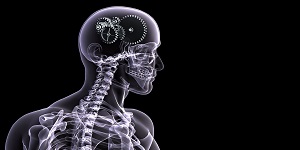
Session 4: Neuro-Imaging
In recent years, brachial plexus magnetic resonance imaging (MRI) has become a safe and accurate way to identify brachial plexus neuropathy in children and adults. Although the clinical distinction between brachial plexus neuropathy and cervical radiculopathy or nerve injury has long been based on nonspecific physical examination and electro diagnostic testing methods, MRN now allows detailed questions about the anatomy and pathology of the nerve. Peripheral, as well as the evaluation of the surrounding soft tissue and muscle tissue, thus promoting an accurate diagnosis. Readers will learn about the current status of brachial plexus MRN, including recent progress and future directions, and will gain insight into brachial plexus neuropathy in adults and children that can be characterized using these techniques.
Radiology Congress | Radiology Congress | Radiology Meetings | Radiology Meetings | Interventional Radiology Workshops | Radiology Events | Trauma & Radiology Conferences | Radio science Conferences
Session 5: Oncology
Radiation therapy and radiation oncology play a key role in the clinical management of patients with tumor diseases. Positron emission tomography (PET) is gaining more and more clinical importance in the management of tumor patients undergoing radiotherapy, because PET allows the visualization and quantification of tumor characteristics at the molecular level, which goes beyond the simple conventional image display. Morphological domain, such as tumor metabolism or receptor expression. Information on tumor metabolism or receptor expression derived from PET can be used as a tool to visualize tumor extent, assess response during and after treatment, predict failure modes, and define the volume that requires augmentation of the dose.
Radiology Congress | Radiology Congress | Radiology Meetings | Radiology Meetings | Interventional Radiology Workshops | Radiology Events | Trauma & Radiology Conferences | Radio science Conferences
Session 6: Interventional Radiology
Interventional Radiologists (IRs) are board-certified radiologists who specialize in minimally invasive targeted therapy. More specifically, they use existing imaging systems, from X-rays to MRI, to advance the catheter through the body (usually in the arteries) to non-surgically treat the source of the disease. According to the Society for Interventional Radiology (SIR), "As the inventor of angioplasty and catheter-placed stents, IR pioneered modern minimally invasive medicine." In many ways, interventional radiologists remain at the center of interventional sports. IR can visualize, open, and stent the carotid and peripheral arteries (including the renal artery, popliteal artery, and femoral artery, etc.). They can help treat aortic aneurysms and dissections, use percutaneous access to treat problems, and use tools such as CT scans, MRIs, PET scans, and ultrasound to view many areas of the body to treat and diagnose patients.
Radiology Congress | Radiology Congress | Radiology Meetings | Radiology Meetings | Interventional Radiology Workshops | Radiology Events | Trauma & Radiology Conferences | Radio science Conferences
Session 7: Cerebral Aneurysm Imaging
Despite advances in treatment, subarachnoid hemorrhage caused by ruptured intracranial aneurysm is a devastating disease, associated with approximately 50% of overall mortality and high survivor morbidity. Neuroimaging is a key factor in the evaluation and treatment of patients with brain aneurysms. Each neuroimaging technology has its unique advantages, disadvantages, and current developments. Two main noninvasive radiological techniques, computed tomography angiography (CTA) and magnetic resonance angiography (MRA), and digital subtraction angiography (DSA) are used to evaluate the efficacy of brain aneurysms. These techniques can comprehensively assess the size, location, rupture status, and other imaging characteristics of aneurysms, and guide clinicians in making appropriate and timely treatment recommendations.
Radiology Congress | Radiology Congress | Radiology Meetings | Radiology Meetings | Interventional Radiology Workshops | Radiology Events | Trauma & Radiology Conferences | Radio science Conferences
Session 8: Abdominal Radiology
The advent of multi-detector array CT (MDCT) has revolutionized many things in abdominal radiology. MDCT is a technique with a variety of application possibilities specific to the abdomen. Imaging of the colon by MDCT has excellent sensitivity for detecting polyps as small as 5 mm, but the visualization of smaller polyps is not yet reliable. Therefore, the debate continues on how to treat patients with microscopic polyps detected or suspected on CT colonography. The risk of malignant transformation of such small polyps is almost nil and it may be reasonable to simply follow these patients. Magnetic resonance imaging has become a widely accepted liver imaging technique. There are several MRI contrast agents for liver imaging that can optimize the study for each indication. Dynamic Small Bowel MRI restores the imaging foundation for patients with small bowel obstruction from computed tomography and barium imaging.
Radiology Congress | Radiology Congress | Radiology Meetings | Radiology Meetings | Interventional Radiology Workshops | Radiology Events | Trauma & Radiology Conferences | Radio science Conferences
Session 9: Emergency and Trauma Care
Multi-slice CT (MCCT) has become an ideal imaging technique due to its high diagnostic accuracy and fast scanning speed, especially in the field of trauma therapy. Since the first introduction of computed tomography, the development of modern computed tomography scanners will inevitably lead to a significant reduction in scanning time, especially in the area of ​​whole-body imaging in trauma room care. The delay time depends on the patient, as it takes time to center and prepare the patient before starting the CT scan. Later is also the time to perform multi-planar reconstruction (MPR) and send the image to a dedicated PACS for reading.
Radiology Congress | Radiology Congress | Radiology Meetings | Radiology Meetings | Interventional Radiology Workshops | Radiology Events | Trauma & Radiology Conferences | Radio science Conferences
Session 10: Clinical Correlations in Radiology
In the field of radiology, some cases require a simplified approach to overcome today's newer medical complications. New complications should be explained and reported in the forum to understand the impressions of other experts. Mainly trauma and cancer treatment are the main research areas of the radiology department, since the new complications come mainly from the aforementioned aspects. Recent complications are not limited to old age, but also affect the younger generation, and even lead to uncertain situations. It takes an hour to discuss the same thing.
Experts from major manufacturing companies have been considered together with other interested parties. This is done to verify and collect key information to assess trends related to the market during the forecast period. Top-down and bottom-up methods have been used to estimate the global and regional scale of this market. Data triangulation techniques and other comparative analysis are also used to calculate the exact size of the global medical image management market.
Market definition assessment and identification of key players and analysis of their strategies to determine the competitive market prospects, opportunities, driving factors, constraints and challenges of the market during the forecast period. A comprehensive quantitative analysis of the industry from 2016 to 2024 enables stakeholders to take advantage of current market opportunities. In-depth analysis of the industry based on market segments, market dynamics, market size, competition, and companies involved in the value chain. Global medical imaging management Conduct comprehensive market analysis and segmentation for products, end users and geographic locations to help with strategic business planning.
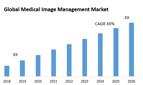
In the growing population, the incidence of chronic diseases is increasing, the preference and demand for minimally invasive surgery is increasing, the level of investment that contributes to the development of advanced technology products is skyrocketing, and the provision of adequate training and safety are Some Factors That May Be Possible Will Promote The Growth Of The Radiology Market During The Forecast Period Of 2020-2027. On the other hand, more and more research and digitization activities will promote even more diverse opportunities, which will lead to the growth of the radiology market during the forecast period mentioned above.
Scope and Importance:
Radiology is a clinical field that is significant and extremely smart to each imaging proficient. There are many captivating perspectives in the field of radiology one such viewpoint is interventional radiology. Interventional radiologists make little entry points, generally in your mid-region, and use needles and catheters to treat conditions inside your body. Clinical pictures are utilized to direct their catheters through your veins, conduits, and organs Interventional radiology lessens cost, recuperation time, agony, and hazard to patients who might some way or another need conventional open a medical procedure. Along these lines, IR has become the essential method to treat numerous sorts of conditions. The medicines IR can successfully perform are always showing signs of change and extending. The absolute most popular IR medicines are angioplasty, stenting, thrombolysis, and embolization.
Radiology Associations in USA:
American Association of Medical Dosimetrists (AAMD), Association for Medical Imaging Management (AHRA), American Society of Nuclear Cardiology, American Society of Radiologic Technologists, American Society for Radiation Oncology, Association of Vascular and Interventional Radiographers, Canadian Association of Medical Radiation Technologists, Radiological Society of North America, Society of Diagnostic Medical Sonography, Society for MR Radiographers and Technologists, Society of Nuclear Medicine and Molecular Imaging.
Radiology Associations in Europe:
Armenian Association of Radiologists, Austrian Roentgen Society, Belarusian Society of Radiologists, Belgian Society of Radiology, Bulgarian Association of Radiology, Association of Radiology of Bosnia & Herzegovina, Croatian Society of Radiology, Danish Society of Radiology, German Radiological Society, Hellenic Radiological Society, Hungarian Society of Radiologists, Irish Institute of Radiography and Radiation Therapy, Italian Society of Radiology, Lithuanian Radiologists’ Association, Norwegian Society of Radiology, Portuguese Society of Radiology and Nuclear Medicine, Polish Medical Society of Radiology, Russian Association of Radiology, Slovak Association of Radiology.
Radiology Associations in Middle East:
Radiology Society of Emirates, Franco-Moroccan Radiology Association, Kuwait Radiological and Imaging Society, Saudi Interventional Radiology Society, Palestinian Association of Medical Radiation Technologists, Algerian Society of Radiology Pan Arab Interventional Radiology Society, Mexican Society of Radiology and Imaging, Oman Radiology and Molecular Imaging Society Turkish Society of Radiology.
Radiology Associations in Asia:
Chinese Society of Radiology, Hong Kong College of Radiologists, Indian Radiological & Imaging Association, Iranian Society of Radiology, Japan Radiological Society, Korean Society of Radiology, Lebanese Society of Radiology, Malaysian College of Radiology, Nepal Radiologists' Association, Sri Lanka College of Radiologists, Taiwan Association of Medical Radiation Technologists, Vietnamese Society of Radiology and Nuclear Medicine, Radiological Society of Thailand
Top Radiology Universities in USA
- Harvard University, USA
- Imperial College London, USA
- Duke University, USA
- Columbia University, USA
- Icahn School of Medicine at Mount Sinai, USA
- University of Toronto, Canada
- Stanford University, USA
- University of Pennsylvania ,USA
- University of Oxford , UK
- University of Washington , USA
Top Cardiology Universities in Asia
-
National University of Singapore , Singapore
-
Tel Aviv University, Israel
-
Seoul National University, South Korea
-
Kyoto University , Japan
-
Capital Medical University, China
-
University of Ulsan, South Korea
-
Yonsei University, South Korea
-
Peking University, China
-
Shanghai Jiao Tong University, China
-
University of Hong Kong , Hong Kong`
Top Cardiology Universities in Europe
-
Université de Paris , France
-
University of Copenhagen, Denmark
-
University of Amsterdam, Netherlands
-
Erasmus University Rotterdam, Netherlands
-
University of Hertfordshire, UK
-
Postgraduate Medicine, England
-
Middlesex University, UK
-
Queen Mary University of London, England
-
Trinity College Dublin, Ireland
-
University of Edinburgh, Scotland
-
Harper Adams University, UK
Top Radiology Hospitals in the World:
-
Stanford Health Care, CA
-
Mayo Clinic, Rochester
-
Cedars-Sinai Medical Center, Los Angeles
-
New York-Presbyterian Hospital-Columbia and Cornell, New York City
-
Massachusetts General Hospital, Boston
-
Mount Sinai Hospital, New York City
-
Brigham and Women's Hospital, Boston
-
University of California, California
-
Los Angeles Medical Center, Los Angeles
-
Stanford Health Care-Stanford Hospital, USA
-
Northwestern Memorial Hospital, Chicago
Meetings International is a global leader in organizing conferences and workshops in all major fields of science, technology and medicine. Meetings International websites are being visited by doctors, engineers, young and budding researchers, entrepreneurs, eminent scientists, academicians and Nobel laureates from various sectors of medical and non-medical sciences. We can target impressions of your advertisements by conferences subject or region and report results in comprehensive detail.
Radiology 2022 attracts different companies and startup businesses to digitally advertise their products and services on its web platform which will help in informing customers quickly and efficiently. The quality and diversity of our advertising options provide clients with the very best customized marketing opportunities. Our advertising platform will provide you is the best chance of showcasing your products/services, and branding your company.
Bring your product to life and create instant brand awareness with a banner ad placement. Make your banner interactive to direct people to where you want them to go. We offer banner ads on our website which will be beneficial for life science professionals across the globe.
Our subscribers and conference attendees can be your upcoming enthusiastic customers. Here is your opportunity to advertise in the website that can connect you to leading experts and specialists worldwide.
Advertisement banner should be provided by the advertising company and must be in jpg or jpeg format. The banner must be of high resolution and must not have copyright infringement.
Any queries regarding advertising opportunities, please contact our representative: contact@meetingsint.com
Meetings International has always been at the forefront to support and encourage the scientific and techno researchers to march forward with their research work. Radiology 2022 conference offers various awards as reorganization to exceptional researcher and their research work. We invite all enthusiastic researchers from all around the world join us for the “2nd International Conference on Radiology” scheduled in Vancouver, Canada during October 19-20, 2022.
Eminent Key Note Speaker Award:
Radiology 2022 will confer the Model Key Note Speaker Award to the researchers who have spent a considerable time of their academic life towards the research related to conference topic. Key note speaker award is for the researchers/speakers who have done all the hard work behind the scene. It will be a small clap for their dedication and hard work.
Outstanding Speaker Award:
Radiology 2022 will bestow model speaker award to those researchers who have made significant contribution towards the conference topic during their research period as well as presented the research topic in an impressive way in the oral presentation during the summit. It will be an apt appreciation from the Jury as well as from the delegates. The award they receive will motivate them to march forward with the research work.
Model Organizing Committee Member Award:
Radiology 2022 will take this opportunity to facilitate eminent experts from this field with the model Organizing Committee Member award for their phenomenal contribution towards the society through their academic research. This will be a trivial but significant reorganization of their dedication and discipline.
Promising Young Researcher Award:
The motive of this Award during the Radiology 2022 is to appreciate research work of the budding young researcher who is continuing their research work to create a better society. The award will provide them a tremendous amount of encouragement to keep moving forward.
Educative Poster Award:
The educative poster award for the Radiology 2022 will be endorsement for those who wishes to display their research paper through a poster and will act as a guiding force those researchers. The award will be presented to the most informative and educative research poster.
Criteria:
-
All presented abstracts will automatically be considered for the Award.
-
All the presentation will be evaluated in the conference venue.
-
All the awards will be selected by the judges of the award category.
-
The winners will be formally announced during the closing ceremony.
-
The winners of the Poster Award will receive award certificate.
The awards will be assessed as far as plan and format, intelligence, argumentation and approach, familiarity with past work, engaging quality, message and primary concerns, parity of content visuals, and by and large impression.
Guidelines:
-
All submissions must be in English.
-
The topic must fit into scientific sessions of the conference.
-
Each individual participant is allowed to submit maximum 2 papers.
-
Abstract must be submitted online as per the given abstract template.
-
Abstracts must be written in Times New Roman and font size will be 12.
-
Abstract must contain title, name, affiliation, country, speaker’s biography, recent photograph, image and reference.
Each poster should be approximately 1x1 M long. The title, contents and the author’s information should be clearly visible from a distance of 1-2 feet.
Meetings International is announcing Young Scientist Awards through “2nd International Conference on Radiology” (Radiology 2022) which is scheduled in Vancouver, Canada during October 19-20, 2022. The conference is mainly focused on cumulative correlations in clinical radiology. Radiology 2022 and upcoming conferences will recognize participants who have significantly added value to the scientific community of Cardiology and provide them outstanding Young Scientist Awards.
The Young Scientist Award will provide a strong professional development opportunity for young researches by meeting experts to exchange and share their experiences at our international conferences. Radiology 2022 aims on the level of thought that individual patients need at various centers in their course. Radiology conference organizing committee conference is providing a platform for all the budding young researchers, young investigators, post-graduate/Master students, PhD. students and trainees to showcase their research and innovation. Eligibility: Young Scientists, faculty members, post-doctoral fellows, PhD scholars and bright Final Year MSc and M.Phil. Candidates. Persons from Scientific Industry can also participate. Benefits: The Young Scientist Feature is a platform to promote young researchers in their respective area by giving them a chance to present their achievements and future perspectives.
-
Acknowledgement as YRF Awardee
-
Promotion on the conference website, Young Researcher Awards and certificates
-
Link on the conference website
-
Recognition on Meetings Int. Award Page
-
Chances to coordinate with partners around the world
-
Research work can be published in the relevant journal without any publication fee.
Criteria:
-
All presented abstracts will automatically be considered for the Award.
-
All the presentation will be evaluated in the conference venue
-
All the awards will be selected by the judges of the award category
-
The winners of the Young Scientist Award will receive award certificate.
The awards will be assessed as far as plan and format, intelligence, argumentation and approach, familiarity with past work, engaging quality, message and primary concerns, parity of content visuals, and by and large impression.
Guidelines:
-
All submissions must be in English.
-
The topic must fit into scientific sessions of the conference
-
Each individual participant is allowed to submit maximum 2 papers
-
Abstract must be submitted online as per the given abstract template
-
Abstracts must be written in Times New Roman and font size will be 12
-
Abstract must contain title, name, affiliation, country, speakers biography, recent photograph, image and reference.
Conditions of Acceptance: To receive the award, the awardee must submit the presentation for which the award is given, for publication at the website, along with author permission. Failure to submit the PPT and permission within the designated timeframe will result in forfeiture of award. Award Announcements: Official announcement of the recipients will occur after the completion of Radiology Conference.
- Cardiac Imaging
- Nuclear Medicine
- Pulmonary Embolism Scanning
- Neurography
- Oncology
- Cerebral Aneurysm Imaging
- Abdominal Radiology
- Interventional Radiology
- Emergency and Trauma
- Clinical Correlations in Radiology
7 Organizing Committee Members
Viroj Wiwanitkit
Professor
Hainan Medical University
China
Benoit Chenais
Professor
Le Mans University
France
Syed Rahmanuddin
Director
3D Cancer Radiology Center
USA
Huiming Yu
Associate chief physician
Peking University Cancer Hospital
China
Mohamad B. Haidar
Director
American University of Beirut Medical Center
Lebanon
Bin Zhu
Director
Nanjing University School of Medicine
China
Faiq Shaikh
Director
University of Pittsburgh Medical Center
USA
2 Renowned Speakers
JUAN ZHANG
Beijing University of Chinese Medicine
China
VLADISLAV TARANOV
Russian National Research Medical University
Russia
Media Partners & Collaborations
Media Partner

Media Partner
Media Partner

Media Partner
Media Partner

fgsg
tgh

Dear Eliza, Thank you for that wonderful opportunity given to share my experiences during the conference. I would like to place on record the smooth conduct of the conference. My appreciation goes a long way to Eliza for her timely communication and well advanced information disseminated processes. The presentation schedule and the time mentioned in the mail sent were also strictly followed. All the best. Regards Kalyani Kenneth Speaker | Psychiatry-2021
Kalyani Kenneth

Dear Laura, Thank you for giving me the opportunity to present my current work! I really enjoyed listening and connecting to a wider international scientist through this conference. And I am really looking forward for the physical conference next year! Best regards Sharmaine
Sharmaine Reintar

Many thanks Laura. It iş my plesure work with you
Pinar Kara

It was my pleasure to be a part of the conference. The talks were technically profound. Thank you once again.
Anupam Mukherjee

Thank you very much for the message. I am glad that the presented poster was appreciated. I'm also grateful for the certificate. Dispite my main work was the subject presented in the session, it would be a pleasure to participate in the webinars.
Ana Rita Domingues

Thank you for your appreciation. I love the support received from you and your admirable patience from invitation till the end of the conference. Thank you very much. I hope to participate in future conferences.
Munezza Khan

Am really grateful for you inviting me to this great event... Am thrilled to be part of this great team, I feel honored 😀😃😄🤝🏻
Aysha Haruna

The conference was arranged nicely through a Webex. I found the sessions interactive and informative. I am looking forward to join the next annual meeting of plant genomics in osaka, japan in april 2022.
Ruchika

Highly appreciate your quick response and I am grateful for all your help.
Dr. P T Sunderam

Informative and Innovative sessions
Michael Lacroix

Delightful event with a wonderful sessions
Mohamed Ebraheem Elmesserey

Very much grateful for this opportunity to share the innovative ideas
Mahmoud Metwaly Taha

It was really informative and excellent platform to share the research ideas
Raktima Chakrabarti

Wonderful experience
KHALED NASR ELDIN REYAD

Thank you very much for organizing this wonderful conference
JIMMY KAYASTHA

it was impressive and left me a very good memory.
VAISHALI SAMIR JOSHI

I learnt a lot from the conference and hope to attend the next conference
Deepak Mane

It was a nice and well-organized meeting! Thank you for all your efforts of having put it together.
Vladimir Startsev

The conference was exciting. I enjoyed so much meeting new colleagues at the venue. I would like to express my sincere gratitude to you for your assistance and opportunity. It was a good experience attending this conference.
Daniela Capdepon

It was great being a part of Surgery 2021
Hadi Nural

How much I appreciate your support, you and your job are meaningful. I always wish the best for you. Thank Laura a lot
Kevin Ton

Hats off to you and fellow organizers. Great opportunities for sharing and networking!
Karen Swanepoel

It was a pleasure to participate in this year’s meeting (even in webinar format due to the COVID-19 pandemic) and I hope the participants enjoyed my talk. There were some interesting talks taking place today and the whole experience was enjoyable like last year. Please rest assured that I will be happy to join next year’s meeting as well, hopefully with a physical presence.
Vasileios Fotopoulos

It was my pleasure to attend this conference. Thanks for your kind efforts.
Saeed Taheri

Thank you for your effort for having made "Pharmacology 2019 conference" wonderful success. Thank you again for that.
Ryong Nam Kim

Thanks for holding a nice conference.
Ramin Ataee

I enjoyed the conference. Italy will be a great place to have the next conference. I look forward to 2020 edition.
Paul Njiruh Nthakanio

I am very happy to make a seminar in the conference. If I have a chance to present my work in your conference, I would talk it in your conference in future.
Takayuki Momma

It was good to connect with a diverse group of scientists - thanks very much for the invite
Thomas P Brutnell

It was a nice experience for me to attend plant genomics conference in Osaka and meet researchers from different countries.
Behnam Derakhshani

It has been an excellent experience.Thank you a lot for this opportunity. I enjoyed it very much!
Julio Cesar Vega

I definitely would like to join your conference again. It was so great about everything. I think I would say great coordination and services.
Maryam Jenabi

Great experience being a part of this conference as a moderator
Soizic de Beaucorps

Amazing experience full of knowledge and meeting new people. Looking forward to being back
Thomas Frederick Hartley

Thanks a lot. It was an excellent event. Really enjoyed.
Umesh Prabhu

It was a pleasure and a great opportunity to be one of the Keynote speakers. I welcome the opportunity to speak at additional conferences in the future. Please keep my contact information for future consideration.
Sharon Nixon-Crenshaw

Thank you for assisting in my participation in a superbly organized Conference. I especially appreciate that you made a special session for me. And once again, sincerely thank you for inviting me to the Conference.
Albert Krashenyuk

Thank you for all your help for the meetings. It was for me a great experience to be in a kind and so scientific group.
Katia lollai

I enjoyed a lot thank you. I thank Meetings International for the quality of the event. All the speakers i had notice of were well prepared and highly motivated Compliments!
Dora Dragoni Dıvrak

Thank you for organizing the conference. It was nice to partake in the event.
Marcello Menapace

I enjoyed participating in the congress and thank you for your efforts to make it easy for participants. Please keep me in mind for future meetings.
Debendra Kumar Tripathy

It was a nice meet and we had a wonderful time in Paris Thank you once again for the arrangement. I would be interested in participating future meetings.
Prasanna Udupi Bidkar

Thank you so much for giving me a chance to my research presentation in your conference.
Seyedataollah montazam

I enjoyed being in the conference. Thanks for you excellent arrangement for my participation. Looking forward for next meetings.
Afaf El Ansary

The conference is very nice, Thank you so much for inviting me to your esteemed event.
Esmira Naftali

The conference was very good. I hope to attend similar meeting in future. Thank you .
Lia Monica Junie

I am indeed glad to acknowledge that for last two days during Optics-2018, International conference on Lasers, Optics and Photonics it was Successfully conducted and we had very fruitful discussions and interactions to make many great friends for life. Many Delegates had interactions with many famous Japanese University Professors and company Managers too for their future possibilities of R&D collaborations. I had arranges a session for such interactions while eating and discussing with relaxed environment and taking group pictures. The hospitality provided by your Organization Committee Members was excellent to help completing Conference Inauguration, all Keynote Speeches, Oral Presentations And Poster session had very high-quality research presentations in many advanced research areas such as Spectroscopy, Ultra high power Lasers, Fibers, Advances in stabilized high frequency mode-locked pulsed Fiber Lasers, MEMS, Optical Interconnects, Photonics and Advanced Optical Biomedical Imaging, THz and optical communications and Interconnects, theoretical and experimental research and so on from highly recognized Professors and Researchers in Japan and advanced institutions in many advanced countries.
Dr. Brahm Pal Singh

I wish to thanks you for my perfect time on conferense in Osaka. It was great experience for me.
Anton Podkopaev

I wish to thanks you for my perfect time on conferense in Osaka. It was great experience for me.
Dr. Anton Podkopaev

I would refer this organization future conferences to my colleagues and also love to join again in diabetes related events. More topics on diabetes should be there in future diabetes conferences.
Radia Boufermes

Thank you for the invitation and the opportunity to participate. It was very good.
Juliana Francisca Grossi Heleno

Thank you very much for the invitation and it was a great honor for me to join this conference. I had a great time.
Alkis Konstantinopoulos

It was a good event and got to make good friends out there.
Nur Ozel

Thank you so much. Really we appreciate the Congress, hope next time it will be longer. Everything was ok, the venue, colleagues, organizing team. Thank you and hope to meet soon in other Congress soon.
Sameh Samy Abdou

It was a great pleasure of mine to be there during the conference looking forward to joining your future conferences
Ahmed Halim Ayoub

The theme of the conference and the scientific panel are very interesting! I am looking excited to learn many new things on this innovative platform
Lisa mattheu

I would love to attend the Infectious Diseases 2019 conference. The hospitality, the renowned speakers, and the city are awesome. Looking forward to it!
Dr. Ianane Jireh Ramos canizares

Hello Daniel, Thank you for the invitation to the conference. It was an interesting experience. I am glad that my presentation was liked. Like the effects of my work. Stay in touch. Best Regards !
Aneta Zymon

Thank you to Daniel Raybin with Wound Care 2018 for allowing me to be the Keynote Speaker for the conference. Dr. Jeff Mayo and I met colleagues from other countries such as Poland and Ukraine that work in plastic medicine and surgery. We are excited to share our knowledge of wound care in the veterinary industry so that our human medicine counterparts can offer the same standardized care that we utilize in the US. Thank you to Regenlabs for sponsoring the event and Jorgensen Laboratories for providing us with a great sponsor to represent our mission.
Nicole LaForest

Dear Daniel! Thank you for the conference, for your accommodation and the opportunity to see the most romantic, mysterious, fabulous Amsterdam
Nadiia Nor

Congress was good, the quality of the presentations was good. The choice of Singapore as strategic headquarters has been good
Buonocore D

Thanks for your invitation. I enjoyed participating at he conference in Rome and am interested in participating again next year in Paris.
Daniel Benetti

Great experience!! Thanks a lot for the opportunity to speak.
Jose Carlos Ferreyra Lopez

Dear Julie, Many thanks to you as you offered me this opportunity to come and participate at this valuable event in Chicago. I think you are great professional and made huge efforts for the event in terms of organization, engaging speakers, etc. And also you have sense for people, act as empathic person what is very important for me. I was very happy and honored to be invited as a speaker at World Biosimilars Conference in terms to present company Ewopharma AG as business partner of different pharma companies in Central and Eastern Europe, especially company Biogen with its portfolio of biosimilars and share my commercial experience in launching biosimilars! I found the conference interested in terms you selected very qualitative speakers from different areas: innovation and science, manufacturing and commercialization. From my perspective, the most interesting topics were: Sarfaraz K Niazi: “Biosimilars: Why are they so widely misunderstood?” Ronald P Dudek: “The adapter CAR Platform: From antibody to CAR T cell therapeutic” Jose Carlos Ferreyra Lopez: “Market access barriers and market value in Mexican public sector for biosimilars” Milind Antani: “Similar biologics in India- Impact of regulations on business” Joel I Osorio: “RegenerAge System “ (So I will be very happy if you could share these mentioned presentations with me!). I can see more potential for further improvement to put 100% focus on Biosimilar topics and to attract more specialists/professionals to gather. It will be excellent if you could attract more speakers from Europe to share their experience. So please consider this option how to attract them in the future (if I got some nice ideas will share with you!).
Sandra Simic

Thanks for giving opportunity to share my research at 3d printing conference .I meet global experts and exchange our ideas.Two days conference are going very good ,workshop session,exhibition .I felt moved to contribute next year also..
Rajkumar Velu

It is wonderful conference of 3d printing & Bio printing in health care .participates are coming from world wide 3d printing & Bio printing experts .Workshop sessions is too good .
Lifeng Kang

• Good workshop session at conference with lots of discussion among audiences and speakers about the regulatory aspect of 3D printed medical devices. • Many of the topics were good and interesting. • It will be good if the organizer could have invited more participants as well as selecting the speakers based on proven and relevant track record. • It will be better if there are people who keep track of the time for each topic.
Albert Sutiono

Conference was superb. It was well conducted. I appreciate all the speakers, presenters and organizers. I am very proud to be part of this event.
Rajani singh

We had a very good session at cardiomersion in which we discuss about the integrated cardiomersion approach to the delivery of cardiovascular case. Thank you to all the speakers who came across the globe. Finally I would like to thank the organizers for making such a remarkable event.
Deepak Puri

I have been a part of this conference and I am very proud to see the conference very well organized and people are helping us and each other to present their case reports and research. Thank you very much to the organizers for making such great events
Suresh Vatsyayan

The event was very nice and I want to thank all the participants, presenters and the organizers. We hope in future we will have more events like this
Yuan Chen

The BABE conference was very interesting and lot of scientific sessions were covered and highlighted. Special thanks to the Organizing Members and the participants
Kateryna Zupanets

Conference was good, thanks for organising. I felt moved to contribute throughout, and felt that to a certain extent I acted as a moderator throughout the event .It is very happy for me.
Alexander M. Korsunsky

I want to express my graduate and thanks to you for all efforts you put to organize such a successful conference. For 2 days I enjoyed the company of brilliant and beautiful minds from all over the world. I had a great chance to exchange expercties with them and in large my horizons. I want to thank you again for organizing the International Conference of Petroleum Engineering 2018, Dubai, UAE and hope to meet you again very soon.
Essa Georges Lwisa

I enjoyed participating in the congress and thank you for your efforts to make it easy for participants. Please keep me in mind for future meetings relating to bariatric surgery, I would be interested in participating or being part of the organizing committee.
Peter Harris

Meetings International meetings are great way to receive way to receive other scientists in different parts of the world doing, These meetings provides the opportunity for you to work at latest concepts from different peoples also specifically it allows the opportunity to call to the new relationships when it resolves in future collaboration across the photolamps
Suzanne Tinsley

I enjoyed conference with Meetings International it is a mutal legitimate.It is always with first move which encourages scientists for practicing ability based practice It also gives lots of opportunity for discussions as well as collaborations in future
Dr. Marie Vazquez Morgan

We enjoyed the meeting so much. Wish you all success in the coming meetings.
Richa Jaiswal

It was a great experience for us to attend the conference. Had good interactions with speakers. Many thanks to you and Mr Peter Harris for giving us opportunity to participate and visit Japan as well. Wish to attend further conferences in future!
Kalpana Kulkarni

The first day was very good. It was meaningful to spend academic meeting. I thank you for your consideration.
Gagan Dhall

International meeting was good looking forward for next meetings.There can be more we can do because there is always scope for improve.
Suman Lata

Thanks for your greeting letter! We enjoyed the conference. Sure! We will meet again in 2019!
Dra Milagrosa C. S. Liu

First of all I would like to say many thanks to YOU and The Organization / Planning Committee, for I have been given the opportunity to join in this very Prestigious Event. I am also grateful to meet with researchers from other countries who have innovative research’s. Hopefully it could upgrade to my knowledge and more increases my interest in this field of science. Nice to join in this event I wish i could be join on the Traditional Medicine 2019.
Yunita Sari Pane

It was great experience for me. My talk was very much liked by all receipients at Osaka.
Mohammad Kamil

I am indeed glad to acknowledge that for last two days during Optics-2018, International conference on Lasers, Optics and Photonics it was Successfully conducted and we had very fruitful discussions and interactions to make many great friends for life. Many Delegates had interactions with many famous Japanese University Professors and company Managers too for their future possibilities of R&D collaborations. I had arranges a session for such interactions while eating and discussing with relaxed environment and taking group pictures. The hospitality provided by your Organization Committee Members was excellent to help completing Conference Inauguration, all Keynote Speeches, Oral Presentations And Poster session had very high-quality research presentations in many advanced research areas such as Spectroscopy, Ultra high power Lasers, Fibers, Advances in stabilized high frequency mode-locked pulsed Fiber Lasers, MEMS, Optical Interconnects, Photonics and Advanced Optical Biomedical Imaging, THz and optical communications and Interconnects, theoretical and experimental research and so on from highly recognized Professors and Researchers in Japan and advanced institutions in many advanced countries.
Dr. Brahm Pal Singh

I wish to thanks you for my perfect time on conferense in Osaka. It was great experience for me.
Dr. Anton Podkopaev

Interesting presentation & worthfull spirit of exchange of experience
Ippei Sakamaki

Thanks a lot we hade a great time and grest conference. We enjoyed the conference .
Chi-Ying Huang

I would like to express my gratitude for your engagement in preparation of the Cancer Therapy Summit 2018. It was a valuable experience.
Hong Qin

Conference was good. Thanks for your support and co operation.
GURUSAMY MARIAPPA

My active partecipation to the Toronto meeting is just over and motivated by it and by Joseph Ndisang who I met there, I'd like to ask and verify whether I could further collaborate more actively, with no expenses for it, within yr network with my long term scientific expertise and long background in the field of diabetes and also at the light of my previous experience with you over the last year.
Marco Songini

“ The organisation and coordination of the international conference of nanotechnology and nanoengineering was at an outstanding level, it was a great honour to participate in such phenomenal event “
Ahmed Abushomi

I also really appreciated all the scientific contact I made within all these participants - Think that in the future if you might request some help for that renewal activity, I might be helping your team of course (I have some suggestions for making better sort / type of presentation activities for invited speakers of course according to my experience in the field).
Jean-paul Lellouche

The conference sesions proceeded successfuly in a hot and friendly atmospher. I observed during the conference every delegate and speaker interacted wit each other and made friend. I am sure that the conference has become a scientific platform to exchange knowledge among the scientists from all over the world, and they wil conduct a new collaborations. During the conference, onsite organizers spent a great efford. I believe that the level and quality and reputation will increase year to year. Unexpected and unavoidable circumstances can occur at everywhere and every activation, and they can be solved easily. Hope to meet you and your team in another conference.
Osman Adiguzel

Thank you for your friendly mail. I enjoyed the conference with interesting speakers. I did not know anybody at the beginning but I found good companions during the conference.The hotel was good but it was located somewhat outside of Paris. Nevertheless, we had a good time. Anyway, thank you again for your invitation.
Nikolaus Stolterfoht

Thank you. Yes the conference was really interesting.
Adil Aghzar

Thank you! I also think that the event was very successful and very interesting. The variety of thematic session and possibility to meet experts from different fields of marine biology and aquaculture was the biggest advantage of the conference. However, it might be useful to attract more participants to next conference.
Magdalena Jakubowska

Thank you for your kind welcome and appreciated support during the appreciable meeting. Thank you for your kind offer to continue to cooperate to this interesting initiative and I am available to cooperate again in remote and to eventually attend the conference. Thank you and your colleagues and the scientific valuable people that attended the conference for your kind cooperation.
Gianluca Ragusa

Thank you for allowing me to participate in this event, I liked the organization and the people who participated, I made many friends too. Of course I would like to collaborate with you in organizing the next Aqua 2019 conference.
Alfredo Olivera Galvez

The meeting was a success with many experience professional.
Nyan Taw

A very thorough and well written set of points. It’s great that you took the time to put this together.
Feng Gao

Thank you for offering an unforgettable experience for all of us. We are honored to attend the conference. We are so happy and thanks a lot.
Stef Stienstra

Many thanks for your continuous support throughout the conference. It was the pleasure to participate and shared the findings at such high level meetings.
Pooja Jain

The conference was very enjoyable and I was honoured to be able to present my research at this prestigious event. The conference was particularly good for me as it is always important to keep up with the new developments
Glenda Gray

I had a wonderful time at the conference and learned so much from the presenters. Thank you kindly for putting this event together.
Marc HV Van Regenmortel

I had a great time in the conference, Everythings are OK, thank you .
Hsiao-Hui Chiu

Wonderful!! thank you for all the help you have done
Sharadha Ramesh

Thank you for your arrangement. Our team enjoyed the meeting so much. Wish you all success in the coming meetings!
CHAN Yui Fung

Surely we will maintain our relation in long run. If there is any opportunity for me to start my carrier being fresher and any guidance from you to me. It will be highly appreciated.
Monika kankarwal

I appreciate your polite contact. I enjoyed my first visit to Singapore. It was meaningful to spend academic meeting. I thank you for your consideration.
Kazue Sawami

Thank you for the email ,it was my sincere honor and pleasure to participate , many thanks for the invitation ,well organised conference, great hospitality ,well composed programme,interesting nursing topics, but not very large group size enough group size to be conducive to excellent discussions, I wish you all the success for your further conference
Hana Kadhom

Thank you for successful conference in Singapore. And thank you for giving me opportunity of oral presentation. It was very good experiences to me.
Hyun, Myung sun.